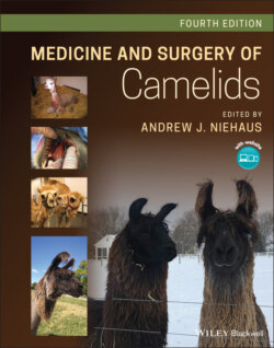
Medicine and Surgery of Camelids

Реклама. ООО «ЛитРес», ИНН: 7719571260.
Оглавление
Группа авторов. Medicine and Surgery of Camelids
Table of Contents
List of Tables
List of Illustrations
Guide
Pages
Medicine and Surgery of Camelids
List of Contributors
Preface
About the Companion Website
1 General Biology and Evolution
Taxonomy
General Biology and Genetics
South American Camelids
Camels
Evolution. Camelid
Camel
South American Camelids
Domestication. Camels
South American Camelids – Llamas and Alpacas
Uses of Camelids
References
2 South American Camelid Behavior and the CAMELIDynamics Approach to Handling
Camelid Behavior
Containment Vs. Restraint
Tips to Low Stress Handling of Llamas and Alpacas
Options for Containment
The Midline Catch, bracelet and Handler Helper
The Neck Wrap – A Thunder Shirt for Camelids
Trimming Toenails
Handling for Veterinary Procedures
No‐Restraint Injections
Box 2.1 Tips for trimming camelids toenails
Box 2.2 Advantages of the No‐restraint Method of Administering Injections
Techniques for No Restraint Injections. Working Alone
Working with an Assistant
Injections for Babies and Weanlings
Drawing Blood
Box 2.3 Tips for Giving Injections
Rectal Palpation and Transrectal Ultrasound
Transabdominal Ultrasound Exams
Physical Exams
Procedures Involving the Head
Oral Medication
Eye Medication
Trimming Fighting Teeth and Incisors
Microchipping
Earring
Sedation
Halter Fit
Box 2.4 Behavioral Problems Related to Improperly Fitting Halters
Box 2.5 Step‐by‐Step Fitting of a Halter
Understanding Male Behavior in Camelids
Pen Size and Shape
Berserk Male Syndrome or Novice Handler Syndrome?
The Novice Handler Syndrome
Further Readings
References
3 Feeding and Nutrition
Anatomy and Nutritional Adaptations
Physiologic Adaptations
Metabolic Adaptations
Camelid Digestive Process
Prehension
Mastication
Saliva
Forestomach Development
Forestomach Fermentation
Forestomach Microbiology
Intestinal Function
Nutrient Requirements of Camelids
Water
Requirement
Assessment
Energy
Maintenance Energy Requirement
Energy Supporting Other Physiologic States
Protein
Maintenance Protein Requirement
Protein to Support Other Physiologic States
Macrominerals
Microminerals
Vitamins
B‐Complex Vitamins
Vitamin A
Vitamin D
Vitamin E
Feeding Behavior and Intake Capacity
Feeding Niches
Dry Matter Intake
Forages and Feed Supplements
Grasses
Legumes
Forbs, Browse and Grass‐like Plants
Commercial Feed Supplements
Mineral Supplements
Feeding Management
Extensive Management
Intensive Management
Dietary Evaluation
Forage Assessment
Feed Label Interpretation
Guaranteed Analysis
Ingredient Listing
Nutrition Related Diseases
Protein‐Energy Malnutrition
Obesity – Hepatic Lipidosis
Forestomach Acidosis
Hypophosphatemic Rickets
Selenium (Se) Deficiency/Toxicity
Copper Deficiency/Toxicity
Zinc‐Responsive Dermatitis
Plant Poisonings
References
4 Physical Exam and Diagnostics
Physical Exam
Conformation
Body Condition and Size
Body Temperature
Cardiac Assessment
Thoracic Cavity Assessment
Abdominal Auscultation
Eye
Ear
Mouth
Diagnostic Procedures. Jugular Venipuncture
Cranial (High‐Neck) Jugular Venipuncture
Mid Cervical Jugular Venipuncture
Low Cervical Jugular Venipuncture
Intravenous Jugular Catheterization
Jugular Venipuncture in Neonatal Crias
Jugular Venipuncture in Camels
Venipuncture in Other Locations
Hemogram and Blood Chemistry
Abdominocentesis
Orogastric Intubation
Nasogastric Intubation
Collection of a Urine Sample
Miscellaneous Procedures. Enema
Thoracocentesis
Bone Marrow Aspiration
Liver Biopsy
Collection of Cerebrospinal Fluid
Radiography
Ultrasonography
Endoscopy
Exploratory Laparotomy and Laparoscopy
Radiographic Imaging
Magnetic Resonance Imaging
Arthrocentesis
Shoulder
Elbow
Carpus
Stifle
Hock
References
Notes
5 Radiology
Introduction
Making Radiographs
Degenerative Joint Disease
Septic Arthritis
Sequestra
Shoulder
Making Skull Radiographs
Tooth‐Root Abscesses
Bulla Disease
Angular Limb Deformities
Patellar Luxation
Rickets
Normal Images
References
Notes
6 Anesthesia and Pain Management
Pre‐operative Assessment. Pre‐Anesthetic Evaluation and Peri‐Anesthetic Period
Sedation
Alpha‐2 Receptor Agonists
Xylazine
Detomidine
Romifidine
Dexmedetomidine
Acepromazine
Benzodiazepines
Diazepam
Midazolam
Field Anesthetic Techniques. Sedation, Tranquilization and Chemical Immobilization
Butorphanol Tartrate
Ketamine Hydrochloride
Tiletamine HCl and Zolazepam HCl (Telazol™)
Muscle Relaxants. Guaifenesin
Injectable Anesthesia
Xylazine‐ketamine
Acepromazine‐Butorphanol‐Tiletamine‐Zolazepam
Dexmedetomidine‐Tiletamine‐Zolazepam
Xylazine‐Tiletamine‐Zolazepam
Propofol
Alfaxalone
Inhalant Anesthetic Techniques. Anesthetic Induction and Intubation
Camel Endotracheal Intubation
Maintenance of Anesthesia Using Inhalant Anesthetics
Agents
Physiology
Monitoring and Supportive Therapy during Anesthesia
Recovery
Anesthetic Complications
Analgesia. Opioids. Fentanyl Patches
Butorphanol
Morphine
Buprenorphine
NSAIDS
Meloxicam
Flunixin Meglumine
Phenylbutazone
Local Anesthesia
Intratesticular and Incisional Line Blocks for Castration
Epidural Anesthesia
Morphine
Lidocaine
Xylazine
Xylazine and Lidocaine
Ketamine
Ketamine and Lidocaine
References
7 Parasitology
External Parasites
Lice
Identification
Life Cycle
Epidemiology
Clinical Signs
Diagnosis
Treatment
Fleas. Identification
Life Cycle
Epidemiology
Clinical Signs
Treatment
Mosquitoes. Identification
Life Cycle
Epidemiology
Clinical Signs
Treatment and Control
Myiasis. Common Flies. Identification
Life Cycle
Epidemiology
Clinical Signs
Management
Black Flies (Buffalo Gnats) Identification
Life Cycle
Epidemiology
Clinical Signs
Treatment
Tabanids (Horse Flies, Deer Flies) Identification and Life Cycle
Epidemiology
Clinical Signs
Treatment
Blow Flies. Identification
Life Cycle
Epidemiology
Clinical Signs
Treatment
Bot Flies. Identification
Oestrus ovis
Camel Nasal Bot Flies
Ticks. Identification
Life Cycle
Epidemiology
Clinical Signs
Tick Paralysis
Mites
Sarcoptic Mange Mite
Psoroptic Mange Mite
Chorioptic Mange Mite
Demodex Mite
Transmission
Diagnosis
Treatment
Internal Parasites. Protozoa
Trypanosomiasis (Surra)
Toxoplasmosis
Coccidiosis
Cryptosporidiosis
Sarcocystosis
Giardiasis
Trematodes
Fascioliasis
Cestodes
Hydatid Disease
Monieziasis
Thysanieziasis
Taenia helicometra, T. hidatigena, T. omissa
Nematodes
Gastrointestinal Nematodes
Etiology and Life Cycle
Epidemiology
Esophagus. Gongylonema spp
Gastric Compartment (C3) Nematodes
Camelostrongylus mentulatus*
Marshallagia marshalli*
Ostertagia ostertagi*
Haemonchus contortus *
Teladorsagia circumcincta*
Trichostrongylus spp.*
Graphinema auchenia*
Spiculopteragia peruvianus*
Small Intestinal Nematodes
Cooperia spp.*
Nematodirus spp.*
Trichostrongylus spp.*
Strongyloides spp
Lamanema chavezi*
Capillaria (Aonchotheca) spp
Bunostomum spp
Large Intestinal Nematodes. Oesophagostomum spp. *
Trichuris tenuis, Trichuris ovis
Skrjabinema ovis
Clinical Signs
Diagnosis. Qualitative Diagnostic Techniques
Box 7.1 Basic Centrifugation Flotation Technique
Quantitative Diagnostic Techniques
Genus‐ and Species‐Level Diagnostic Techniques for Strongyles
Anthelmintic‐Related Diagnostic Techniques
Box 7.2 Steps to Perform the Fecal Egg Count Reduction Test (FECRT)
Post‐Mortem Examination
Treatment and Control Programs
Prevention
Monitoring
Treatment
Biosecurity and Quarantine
Refugia
Sustainable Anthelmintic Use
Combination Anthelmintic Use
Alternative Control Measures
Nematodes of Other Body Systems. Lungworms. Dictyocaulus filarial, D. viviparus, Dictyocaulus spp
Angiostrongylus cantonensis
Brainworm or Meningeal Worm
Eye Worm. Thelazia californiensis, Thelazia spp
References
8 Multisystem Disorders
Neoplasia
Stress
Hyperthermia. Thermoregulation
Predisposing Factors
Clinical Findings
Necropsy Findings
Therapy
Prevention
Hypothermia
Fluid Therapy in Llamas and Alpacas
Failure of Predicted Growth and Weight Loss
References
9 Integumentary System
Normal Camelid Skin
Hair (Fiber, Wool)
Hair Quality
Hair Loss
Skin Glands. Adnexal Glands
Metatarsal Glands
Interdigital Glands
Poll Glands of Camels
Diagnosis of Dermatologic Conditions
Diseases of the Integument. Definition of Terms
Parasitic Diseases of the Skin
Viral diseases of the skin. Camelpox
Etiology
Epidemiology
Clinical Signs
Diagnosis
Management
Contagious Ecthyma
Epidemiology
Clinical Signs
Diagnosis
Treatment
Prevention
Foot‐and‐Mouth Disease
Etiology
Epidemiology
Clinical Signs
Diagnosis
Pathology
Treatment
Prevention
Papillomatosis
Etiology
Epidemiology
Clinical Signs
Diagnosis
Management
Vesicular Stomatitis
Epidemiology
Clinical Signs
Bluetongue
Etiology
Epidemiology
Clinical Signs
Diagnosis
Treatment
Prevention
Fungal Infections. Dermatophytosis (Ringworm)
Etiology
Clinical Signs
Epidemiology
Pathology
Diagnosis
Treatment
Coccidioidomycosis
Epidemiology
Clinical Signs and Pathology
Diagnosis
Treatment
Prevention
Candidiasis
Diagnosis
Treatment
Prevention
Miscellaneous Fungal Infections
Bacterial Infections. Abscesses
Etiology
Epidemiology
Clinical Signs
Diagnosis
Management
Pseudotuberculosis (Caseous Lymphadenitis – CLA)
Etiology
Epidemiology
Clinical Signs and Treatment
Control
Folliculitis and furunculosis (S. aureus dermatitis, botryomycosis, pyoderma, contagious skin necrosis) Etiology and Clinical Signs
Treatment and control
Dermatophilosis (streptothricosis, rain scald, rain rot) Etiology and Epidemiology
Clinical Signs and Treatment
Nutritional Skin Diseases. Zinc‐Responsive Dermatosis (Idiopathic Hyperkeratotic Dermatosis)
Miscellaneous Dermatoses. Follicular/Sebaceous Gland Cysts
Idiopathic Nasal/Perioral Hyperkeratotic Dermatosis (Munge)
Clinical Signs
Diagnosis
Treatment
Idiopathic Neutrophilic/Necrolytic/Hyperkeratotic Dermatosis (Generalized Munge)
Clinical Signs
Diagnosis
Therapy
Burns (Photosensitization, actinic dermatitis, solar dermatitis)
Clinical Signs
Management
Ichthyosis
Pemphigus Vulgaris
Insect bite hypersensitivity (Idiopathic urticaria)
Adverse Drug Reaction
Neoplastic and non‐neoplastic tumors
The Foot. The Normal Foot
Foot Diseases of Camelids. Diseases of the toenail
Pododermatitis, Interdigital Dermatitis and Traumatic Injury
Clinical Signs
Diagnosis
Treatment
Prevention
Mammary Gland. Mammary Gland Anatomy
Milk Production
Milk Composition
Teats
Mastitis
Predisposing Factors
Etiology
Clinical Signs
Diagnosis
Therapy
Prevention
Udder edema
The Ear. Normal Anatomy
Diagnostic Procedures
Diseases. Lacerations
Other Diseases of the Pinna
Otitis Externa
Otitis Media and Interna
Miscellaneous Conditions
Wound Healing in Camelids
Phases of Wound Healing. Hemostasis
Inflammatory (Debridement) Phase
Proliferative (Repair) Phase
Remodeling (Maturation) Phase
Wound Evaluation
Wound Management. Primary, Delayed Primary, & Second Intention Healing
Wound Cleansing
Local Wound Therapy
Wound Dressings
Notes
References
10 Musculoskeletal System
Anatomy and Conformation
Thoracic Limb
Pelvic Limb
Gaits
Specific Conditions. Long Bone Fracture
Principles of Fracture Repair
External Stabilization
Splints and Casts
Thomas Splints
External Fixators and Transfixation Pin Casts
Open Reduction and Internal Fixation
Specific Limb Fractures
Humerus
Radius/Ulna
Metacarpus/Metatarsus
Femur
Tibia
Complications
Vertebral Fractures. Etiology
Clinical Signs
Diagnosis
Management
Fractures in Camels
Mandibular Fractures
Anatomy
Management
Disorders of Joints. Fetlock Hyperextension
Scapulohumeral Luxation. Anatomy
Etiology
Diagnosis
Treatment. Closed Reduction
Open Reduction and Stabilization
Scapulohumeral Arthrodesis
Disorders of the Stifle. Anatomy
Cranial Cruciate Ligament Rupture. Etiology and Clinical Signs
Diagnosis
Treatment
Luxating Patella. Etiology and Clinical Signs
Diagnosis
Treatment
Upward Fixation of the Patella
Diagnosis
Treatment
Tibiotarsal Luxation
Bone Sequestra
Clinical Signs
Diagnosis
Surgery
Juvenile/Congenital Limb deformity
Treatment
References
11 Respiratory System
Overview of the Camelid Respiratory System. Nostrils and Nasal Cavity
Nasolacrimal Duct
Sinuses
Lungs, Trachea and Bronchi
Physiology
SAC Adaptations to Altitude
Diagnostic Procedures
Infectious Diseases
Bacterial Pneumonia
Etiology
Hypertrophic Osteopathy
Parasitic Diseases
Congenital Disorders
Miscellaneous Diseases [75–77] Trauma
References
12 Digestive System and Abdomen
Anatomy and Physiology
Peritoneum, Omentum, and Mesentery
Lips and Oropharynx
Esophagus
Teeth. Comparative Dental Anatomy
Incisors
Canines
Cheek Teeth
Aging
Salivary Glands
Gastrointestinal Tract
Stomach
Gastric Motility
Intestine. Small Intestine [45–47]
Large Intestine [46]
Liver
Bile Duct
Clinical Signs Associated with Digestive Disorders
Anorexia
Dysphagia
Regurgitation and Emesis
Abdominal Distention
Diarrhea
Constipation
Ileus
C1 Atony
Colic
Diseases of the Digestive System. Lip and Tongue Disorders
Stomatitis
Etiology
Signs
Diagnosis
Treatment
Oral Infections. Etiology
Dental and Boney Disease. Dental Disease and Dental Care
Tooth root abscessation and Osteomyelitis and Sequestra
Cheek Tooth Extraction
Retained Deciduous Teeth
Teeth Trimming and Routine Dental Care
Trimming Incisors to Prevent Tooth Drift
Salivary Gland Disorders. Etiology
Signs
Diagnosis
Treatment
Diseases of the Pharynx and Esophagus. Pharyngitis and Esophagitis. Etiology
Diagnosis
Treatment
Esophageal Obstruction (Choke) Etiology
Signs
Diagnosis
Treatment and Prevention
Megaesophagus (Esophageal Dilatation, Esophageal Paralysis and Esophageal Achalasia, Cardiospasm)
Etiology
Signs
Diagnosis
Treatment
Gastric Disorders
Gastric Ulcers
Forestomach (C1) Acidosis (Grain Overload)
Intestinal Disorders
Enteritis
Obstruction
Gastrointestinal Bezoars
Mineral Concretions
Intestinal Ulceration
Rectal Prolapse
Rectal Laceration
Diseases of the Abdominal Cavity. Peritonitis
Intraabdominal Hemorrhage
Diseases of the Liver. Hepatic Insufficiency
Hepatic Lipidosis
Abdominal and Gastrointestinal Surgery. Surgical Anatomy
Patient Positioning
Presurgical Fasting
Anesthesia
Approaches to the Abdomen. Ventral Midline
Flank
Parainguinal
Paracostal
Herniorrhaphy
C1 Gastrotomy
Gastrostomy (Permanent C1 Fistulation)
Surgery of the Spiral Colon
Rectal Prolapse
Rectal Laceration
Atresia Ani and Atresia Coli
Notes
References
13 Endocrine System
Endocrine Organs. Pituitary Gland
Thyroid Gland
Parathyroid Glands
Adrenal Glands
Pancreas
Gonads
Endocrine Disorders. Physiology of Energy Metabolism in South American Camelids
Ketosis, Hyperlipidemia, and Hepatic Lipidosis
Diabetes Mellitus
Hyperosmolar Syndrome
References
14 Hematology, Clinical Biochemistry, and Fluid Analysis
CBC
Erythrocytes
Leukocytes
Platelets and Coagulation
Hematopoietic Neoplasia
Bone Marrow
Flow Cytometry and Immunohistochemistry
Immunodeficiency Disorder in Juvenile Llamas
Biochemistry
Peritoneal and Pleural Fluid Analysis
Cerebrospinal Fluid (CSF) Analysis
References
15 Cardiovascular System
Anatomy and Physiology
Special Diagnostic Procedures. Physical Exam
Other Testing
Diseases
Congenital Heart Diseases
Acquired Heart Diseases. Nutritional Muscular Dystrophy
Infectious Diseases
Toxins
Pericardial Disease
Cardiac Neoplasia
Cardiomyopathy
Arrhythmias
References
16 Reproduction and the Reproductive System
Reproductive Strategies. Vicuñas
Guanacos
Wild Bactrian Camels
Normal Reproduction. Male Llamas and Alpacas. Anatomy
Physiology
Testicular Function
Sexual Behavior
Female Llamas and Alpacas. Anatomy [28–31]
Puberty
Physiology
Pregnancy
Fetal Membranes and Fluid
Pregnancy Determination
Behavior
Hormone Analysis
Rectal Palpation
Ballottement
Ultrasonography
Parturition
Involution of the Uterus [79]
Camel Reproduction. Female Camel Reproduction [41, 80]
Camel Pregnancy
Postpartum Complications
Vaginal Prolapse. Causes
Clinical Signs
Prolapse of the Uterus. Causes
Clinical Signs
Rupture of the Uterus. Causes
Clinical Signs
Uterine Infection
Agalactia
Rejection of the Cria
Obstetric Procedures [85]
Manipulation of Dystocias
Uterine Torsion Correction
Uterine Inertia
Cesarean Section
Pregnancy Termination
Infertility [91] Female Llamas and Alpacas
Etiology
Abortion
Infertility Examination
Treatment of Infertility and Metritis
Infertility Conditions of Female Camels
Male Llamas and Alpacas. Etiology
Infertility Examination
Assisted Reproductive Techniques [17, 25, 117, 118]
Embryo Transfer
Selection of Breeding Camelids
Reproductive Surgery. Cesarean Section
Ovariohysterectomy
Persistent or Imperforate Hymen
Castration [134]
Early Castration – Pro or con
Cryptorchid Castration [27]
Vasectomy
Acknowledgments
Notes
References
17 Urinary System
Anatomy. Kidney
Ureters, Urinary Bladder, and Urethra
Characteristics of Camelid Urine
Diagnostic Procedures
Ultrasonography
Cystoscopy
Urination Behavior
Diseases
Stranguria and Dysuria
Nephrosis
Oak Toxicity
Clinical Signs and Laboratory Findings
Treatment
Gentamicin Toxicity
Clinical Signs and Laboratory Findings
Vitamin D Toxicity
Chronic Renal Failure
Nephritis
Cystitis and Urethritis
Posthitis
Concretions of the Urinary Tract
Clinical Signs
Diagnosis
Treatment
Neoplasia
Congenital Defects
References
18 Ophthalmology
Anatomy of the Eye
Ophthalmic Diagnostic Procedures
Diseases of the Eye
References
19 Nervous System
Diagnosis
Common Neurologic Diseases. Polioencephalomalacia
Meningeal Worm
Encephalitic Listeriosis and Otitis Media/Interna
Tick Paralysis
Less Common Neurologic Diseases. Viral Encephalitides
Tetanus
Botulism
Dysautonomia
Other Reported Neurologic Diseases
Non‐specific Therapy Plan for Neurologic Disease
References
20 Neonatology
Prepartum Care
Characteristics of the Camelid Neonate
Immediate Care of the Newborn
Meconium
Care of the Umbilical Cord
Care Given to the Dam
Prematurity
Signs of Prematurity
Immunoglobulins
Production of Immunoglobulins
Colostrum Composition and Absorption
Failure of Passive Transfer (FPT) of Immunoglobulins
Detection of FPT
Management of FPT
Treatment of FPT
Caring for the Orphaned Camelid
Milk Replacement
Commercial Milk Replacers
Weaning
Neonatal Diseases and Conditions. Umbilical Infection and Associated Complications. Acute Umbilical Infection
Chronic Infection
Umbilical Hernias
Patent Urachus
Osseous Sequestration
Neonatal Septicemia
Hyperosmolar Syndrome
Diarrhea
Viral Diarrhea
Bacteria Diarrhea
Parasitic Diarrhea
Nasolacrimal (NL) Duct Occlusion/Atresia
Neonatal Maladjustment (Dummy Syndrome)
Limb Deformities and Rickets
Congenital Defects (Partial List)
Immunoprophylaxis
Routine Husbandry Practices
Acknowledgments
References
21 Congenital/Hereditary Conditions
Terminology
Management of Congenital/Heritable Conditions
Teratogenesis. Etiology
Hereditary Traits
Chromosomal Aberration
Congenital Conditions
Skeletal Defects. Angular Limb Deformities
Arthrogryposis
Overextension of the Carpus
Shortening of Long Bones
Luxation of the Patella
Hemivertebrae
Polydactyly/Syndactyly
Scoliosis [45, 52, 53]
Luxation of the Tibiotarsal Bone
Face and Head Defects. Craniofacial Dysgenesis
Jaw Dysgenesis [43]
Dysgenesis of the Palate
Reproductive System Defects
Hypogenesis/Agenesis of Reproductive Organs
Intersex
Male Defects
Digestive Tract Defects
Cardiovascular Defects [12, 73]
Miscellaneous Defects [15, 20,76–78]
Nonanatomic Defects
New Congenital Defects of SACs
Camelid Hybrids
Notes
References
22 Toxicology
Adaptation to Toxicants
Diagnosis of Poisoning
Prevention of Poisoning
Treatment of Poisoning
Antidotes
Specific Antidotes
Specific Toxins. Pesticides. Insecticides
Rodenticides
Drug Toxicities and Adverse Drug Reactions
Tolazoline Intoxication
Albendazole
Propylene Glycol
Vitamin D Toxicosis
Growth Promoting Antimicrobials
Prostaglandin Toxicity
Heavy Metals
Selenium
Copper
Toxic Plants [2, 82]
Rhododendron Poisoning
Oleander Poisoning
Pyrrolizidine Alkaloid Poisoning
Dieffenbachia Poisoning (Dumbcane)
Yew Poisoning
False Hellebore Poisoning
Castor Bean Poisoning
Nitrate (Nitrite) Poisoning
Cyanide Poisoning
Iphiona aucheri (Boiss)
Capparis tomentosa (Laturdei)
Trema tomentosa (Poison Peach)
Cyanobacteria
Sodium Fluoride
Mycotoxins
Sporodesmiomycosis (Facial Eczema)
Aflatoxicosis
Ryegrass (Ergot) Toxicosis
Phalaris Staggers
Fluroacetate Poisoning
Fescue Poisoning
Miscellaneous Plant Poisonings
Acorn Poisoning (Tannin Toxicity)
Mechanically Injurious Plants
Foxtails (Hordeum spp.)
Bristle Grass
Tarweed (Hemazonia spp.)
Snake Envenomation
Miscellaneous Bites and Stings [4]
Blister Beetle Toxicity
Notes
References
23 Old World Camelids
Behavior and Handling
Body Posture
Vocalization
Offensive and Defensive Behaviors
Spitting
Biting
Kicking
Restraint
Physical Restraint
Head Control
Tethering
Hobbles
Sedation
α‐2 Agonists
Routine Veterinary Care. Physical Examination & Normal Parameters
Vaccination
Clostridial Diseases
Rabies
West Nile Virus
Bovine Viral Diarrhea Virus (BVDV)
Equine Herpes Virus‐1
Middle Eastern Respiratory Syndrome
Anthrax
Blood Collection
Fecal Examination and Deworming
Reproduction. Normal Reproductive Behavior and Physiology
Assisted Reproductive Technology
Hybridization
Pregnancy Diagnosis
Behavior
Rectal Palpation
Ultrasound
Progesterone
Cuboni Reaction
Pregnancy and Parturition
Neonatal Care
Diseases of Camels. Metabolic Bone Disease
Nutritional Secondary Hyperparathyroidism
Rickets
Sway Disease
Viral Diseases. Middle Eastern Respiratory Syndrome
Foot and Mouth Disease
Bovine Viral Diarrhea Virus
Bacterial Diseases. Caseous Lymphadenitis
Streptococus Equi Subspecies Zooepidemicus
Parasitism
Surgery of Camels
Gastrointestinal Surgery
Cesarean Section
Castration
Dulaa Surgery
Clinical Signs
References
Index. a
b
c
d
e
f
g
h
i
j
k
l
m
n
o
p
r
s
t
u
v
w
x
y
z
WILEY END USER LICENSE AGREEMENT
Отрывок из книги
Fourth Edition
.....
Figure 2.34 In this photo, a helper is balancing the animal for an IM injection.
Figure 2.35 This photo illustrates using a handler helper, along with a balancing hand under the jaw, as the alpaca receives a subQ injection. Notice that the handler injecting the medication is using the wool to lift the skin away from the body, distracting the animal from the entry of the needle.
.....