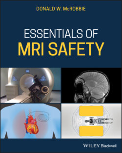
Essentials of MRI Safety

Реклама. ООО «ЛитРес», ИНН: 7719571260.
Оглавление
Donald W. McRobbie. Essentials of MRI Safety
Table of Contents
List of Tables
List of Illustrations
Guide
Pages
Essentials of MRI Safety
Foreword: essentials
Acknowledgments
1 Systems and safety: MR hardware and fields. INTRODUCTION
OVERVIEW OF MRI OPERATION
Nuclear magnetic resonance
Example 1.1 B1 amplitude
Image formation
Slice selection
In‐plane localization
Pulse sequences
Parallel imaging
Overview of MRI applications
MRI HARDWARE
Magnet system
Superconductivity
Superconducting MR magnets
Short larger‐bore magnets
Other magnets
Imaging gradients subsystem
Example 1.2 Gradient performance
Radiofrequency subsystem
RF transmission
RF reception
ELECTROMAGNETIC FIELDS
Static field. Definition of magnetic flux density and the tesla
MYTHBUSTER:
B0 fringe field
Fringe field spatial gradient
MYTHBUSTER:
The imaging gradients
Example 1.3 Bz from a gradient
Example 1.4 Gradient dB/dt
Radiofrequency field
MYTHBUSTER:
B1+ and B1+rms
Scanning modes
OTHER MEDICAL DEVICES
CONCLUSIONS
Revision Questions
References
Further reading and resources
Notes
2 Let’s get physical: fields and forces. BASIC LAWS OF MAGNETISM
Understanding Maxwell’s Equations
Electrical charge and electric fields
Magnetic fields
Electromagnetic induction
Electromagnetic waves
Generating magnetic fields
B field from a long straight conductor
B field from loop conductors
B field from a solenoidal coil
B field from a shielded MRI magnet
Spatial dependence of magnetic fields
MAGNETIC MATERIALS
Ferromagnetism
Demagnetizing field and factors
MYTHBUSTER:
Example 2.1 Magnetization of a nickel coin
Example 2.2 Iron rod in the fringe field
FORCES AND TORQUE
Translational force: non‐ferromagnetic materials
MYTHBUSTER:
Example 2.3 Force on a diamagnetic object
Translational force: ferromagnetic objects
Force on a soft unsaturated ferromagnetic material
Example 2.4 Force on an unsaturated ferromagnetic object
MYTHBUSTER:
Soft saturated ferromagnetic material
Permanent magnet
Example 2.5 Force on a ferromagnetic object aligned with B0
Example 2.6 Force on a ferromagnetic object side on to B0
Projectile velocity
Torque
Torque on diamagnetic and paramagnetic objects
Example 2.7 Torque on a weakly ferromagnetic magnetic implant
MYTHBUSTER:
Torque on soft ferromagnetic objects
MYTHBUSTER:
Example 2.8 Torque on a ferromagnetic cylinder
Torque v translational force
Forces on circuits
Force on a straight conductor
Example 2.9 Force on an electrical wire
LORENTZ AND HYDRODYNAMIC FORCES
Lorentz force
Magneto‐hydrodynamic effect
LAWS OF INDUCTION
Faraday induction from the gradients
Induced fields from movement within the static fringe field gradient
Example 2.10 Movement in the fringe field gradient
Lenz’s law
Induction from the radiofrequency exposure
Wave‐like behavior of B1
Near and far field
MYTHBUSTER:
λ/2 resonant length
MYTHBUSTER:
Example 2.11 RF wavelength at 1.5 T
CONCLUSIONS
Revision Questions
References
Further reading and resources
Notes
3 Bio‐effects 1: static field. INTRODUCTION
PHYSICAL MECHANISMS
Magneto‐hydrodynamic (Hall) effect
Lorentz force
Magneto‐mechanical forces and torque
Example 3.1 Force on an electrolyte ion
Example 3.2 Forces on blood cells
Example 3.3 Torque on a blood cell
Induced electric fields
Example 3.4 Faraday induction from motion
CELLULAR EFFECTS
ANIMAL EFFECTS
EPIDEMIOLOGY
HUMAN PHYSIOLOGICAL EFFECTS
ACUTE SENSORY EFFECTS
Metallic taste
Vertigo and nystagmus
MYTHBUSTER:
Nausea
COGNITIVE EFFECTS
STATIC FIELD EXPOSURE LIMITS
CONCLUSIONS
Revision questions
References
Further reading and resources
4 Bio‐effects 2: time‐varying gradient fields. INTRODUCTION
PHYSICAL INTERACTION
Example 4.1 Electric field induced from a gradient pulse
Example 4.2 Induction in an ellipse
ELF TIME‐VARYING MAGNETIC FIELD EFFECTS
Cellular effects
Do electromagnetic fields in the frequency range 50–60 Hz cause cancer?
Therapeutic magnetic stimulation. Bone healing
Transcranial Magnetic Stimulation
MAGNETIC STIMULATION. Magneto‐phosphenes
MYTHBUSTER:
Example 4.3 Magneto‐phosphene threshold
Nerve and muscular stimulation
Basic electrophysiology of nerves
Forms of the strength‐duration curve
Properties of magnetic stimulation
B‐field change step size
MYTHBUSTER:
Strong stimuli
Waveform dependence
Very short stimuli
Respiratory and cardiac stimulation
Example 4.4 Induction in the heart
PERIPHERAL NERVE STIMULATION IN MRI
Predicting and avoiding PNS
Example 4.5 PNS predicition
EXPOSURE LIMITS
Example 4.6 Cardiac stimulation
CONCLUSIONS
Revision questions
References
Further reading and resources
5 Bio‐effects 3: radio‐frequency fields. INTRODUCTION
PHYSICAL INTERACTION
Radiofrequency in MRI
Specific Absorption Rate
Example 5.1 SAR from linear and quadrature transmission
SAR hotspots
B1 non‐uniformity
MYTHBUSTER:
TISSUE HEATING
SAR and temperature rise without cooling
Example 5.2 Heating of the lens
Temperature rise with perfusion cooling
Other cooling mechanisms
Thermal conduction
Radiative cooling
Other cooling mechanisms
Example 5.3 Radiative cooling
Thermal regulation
BIOLOGICAL EFFECTS
Cellular studies
Animal studies
CEM43
Example 5.5 CEM43 and time to cause a skin burn
Carcinogenic effects
Human studies and epidemiology
Do mobile phones cause cancer?
Microwave hearing
RF burns in MRI
Leads, electrodes, and fixation
Coil faults
Contact with or proximity to the bore
For no apparent reason
Avoiding RF burns
RF EXPOSURE LIMITS
Temperature and SAR Limits
Temperature limitation
SAR limits
Example 5.5 Partial body SAR
Specific Energy Dose
Example 5.6 SED for an examination
Other limits
CONTROLLING SAR IN PRACTICE
Flip angle
RF pulse type
MYTHBUSTER:
Example 5.7 SAR reduction strategies
Number of echoes / number of slices
Changing TR
Hyperechoes
Preparation and restoration pulses
Scan time and delay
Parameters which do not affect SAR
MYTHBUSTER:
Getting a feel for SAR
Example 5.8 SAR exercise
Controlling B1+RMS
Example 5.9 SAR and B1+RMS
Monitoring SAR
Patient registration
What happens if you enter the wrong patient weight?
Example 5.10 Wrong patient weight
SAR prediction and measurement
CONCLUSIONS
Revision questions
References
Further reading and resources
6 Acoustic noise. INTRODUCTION
GENERATION OF ACOUSTIC NOISE IN MRI
Example 6.1 Force on a gradient coil
MEASURING NOISE: dB(A), dB(C), dB(Z)
Sound intensity and pressure level
MYTHBUSTER:
Example 6.2 Combined noise sources
SPL weightings
dB(Z)‐weighting
dB(A)‐weighting
dB(C)‐weighting
Time‐varying noise measurement
Frequency specific noise measurement
Measuring scanner noise
ANATOMY AND PHYSIOLOGY OF HUMAN HEARING
The auditory system
Hearing damage
MRI NOISE EXPOSURE. Field strength and gradient dependence
MYTHBUSTER:
Pulse sequence dependence
Peak frequencies
Scanner design
Scanner noise in the MR room
REDUCING ACOUSTIC NOISE IN PRACTICE
Slice width
Low SAR RF pulses
Field of view
Pixel size
TE and receive bandwidth
TR
Number of slices, echoes
b‐value
Sequence choice
HEARING PROTECTION
Specification of acoustic attenuation devices
The NRR method
MYTHBUSTER:
Example 6.3 NRR and earplugs
The SNR method
The H‐M‐L method
Example 6.4 Protection using HML
The Octave band method
SLC80 method
Hearing protection in practice
Ear plugs
Ear defenders
Limitations of hearing protection
ACOUSTIC NOISE LIMITS
Patient limits
Occupational exposure limits
Example 6.5 Interventional MRI
CONCLUSIONS
Revision questions
References
Further reading and resources
Note
7 Pregnancy. INTRODUCTION
CELLULAR EFFECTS AND ANIMAL STUDIES. Static field
Time‐varying magnetic fields
HUMAN STUDIES AND EPIDEMIOLOGY
Acoustic noise
GADOLINIUM‐BASED CONTRAST AGENTS
MYTHBUSTER:
EXPOSURE LIMITS AND GUIDANCE
MYTHBUSTER:
Fetal SAR and temperature
MYTHBUSTER:
Minimizing SAR
Acoustic noise
Professional guidance
Staff exposure
CONCLUSIONS
Revision questions
References
Further reading and resources
Notes
8 Contrast agents. INTRODUCTION
PHYSICAL AND CHEMICAL PROPERTIES
Example 8.1 Moles and mass
Relaxation properties
Example 8.2 Relaxation times
Physical properties of CGCAs
MYTHBUSTER:
Example 8.3 Number of Gd ions
Example 8.4 Number of Gd ions
CONTRAST REACTIONS AND ADVERSE EVENTS
General reactions
NSF
Retention
Current advice
PREGNANCY AND LACTATION. Pregnancy
Breast‐feeding
CONCLUSIONS
Revision questions
References
Further reading and resources
National guidance documents. Australia
Canada
European Union
New Zealand
UK
USA
Note
9 Passive implants. INTRODUCTION
RISKS FROM PASSIVE IMPLANTS
Static magnetic forces
Diamagnetic materials
Paramagnetic materials
Example 9.1 Torque on a paramagnetic stent
Example 9.2 Translation force on a paramagnetic stent
Ferromagnetic materials
MYTHBUSTER:
Example 9.3 Ferromagnetic aneurysm clip
“Weakly ferromagnetic” materials
Example 9.4 Saturation of stainless steel
Magnetic forces due to motion
Example 9.5 Lenz force on an orthopaedic implant
Example 9.6 Torque from Lenz’s effect
MYTHBUSTER:
Induction
Induced electric fields
Example 9.7 Induced E in a hip implant
Example 9.8 dB/dt from the RF transmission
Vibration
MYTHBUSTER:
Implant heating
Heating by the gradients
Example 9.9 Power dissipation from the gradients
Example 9.10 Metal object suspended in air
RF heating
Example 9.11 Power dissipation from B1
Example 9.12 Implant heating from B1
MYTHBUSTER:
MYTHBUSTER:
MYTHBUSTER:
Electromagnetic and thermal modelling
MYTHBUSTER:
ASTM TESTING
Translational force: ASTM F2502
Limitations of the deflection test
Torque: ASTM F2213
Suspension method
Low friction surface method
Calculation based upon measured displacement force
Radiofrequency heating of implants ASTM F2182
Limitations of the RF heating test
EXAMPLES OF PASSIVE IMPLANTS. Aneurysm clips
Orthopedic implants
External fixation devices
Spinal rods and fixation devices
Stents, coils, and filters
Heart valves and annuloplasty rings
Guidewires, catheters, and leads
Medicinal patches
Other devices
Tattoos, piercings, cosmetics, and clothing
ARTEFACTS
Cause of artefacts
Ferromagnetic objects
Testing for artefacts: ASTM‐F2119‐07
CONCLUSIONS
Revision questions
References
Further reading and resources
Notes
10 Active implants. INTRODUCTION
RISKS FROM ACTIVE IMPLANTS
MRI accidents involving active devices
Static magnetic forces
Effect on reed switches
Forces on leads
Example 10.1 Lorentz force on an ICD lead
Induction from gradients’ dB/dt
Example 10.2 Pacemaker lead induced voltage from the gradients
Induction from the RF dB/dt
Example 10.3 RF induction in a lead
Measurement of lead tip heating. Phantom and in‐vitro measurements
Abandoned and broken leads
Lead configuration
In‐vivo temperature measurements
Example 10.4 SAR and scar tissue
PACEMAKERS AND ICDS
MR conditional pacemakers and ICDs
Scanning procedure
Complying with the conditions
Contraindications
“Legacy” pacemakers
NEUROSTIMULATORS
Deep brain stimulator (DBS)
Example 10.5 DBS electrode
B1+RMS
MR conditions
Specific conditions
MYTHBUSTER:
Other neurostimulators
Vagus nerve stimulators (VNS)
Spinal Cord Stimulators (SCS)
Sacral nerve stimulators (SNS)
Gastric electro‐stimulators (GES)
COCHLEAR IMPLANTS
MR conditional cochlear implants
Adverse events
Example 10.6 Cochlear implant magnet
ENDOSCOPIC CAMERAS
IMPLANTABLE INFUSION PUMPS
Adverse events
MYTHBUSTER:
KEEPING WITHIN THE CONDITIONS
Static field and static field spatial gradient
Gradient slew rate and dB/dt
SAR and B1+RMS
MYTHBUSTER:
The fixed parameter option
Active implant scanning policy
CONCLUSIONS
Revision questions
References
Further reading and resources
Manufacturers’ technical manuals and MRI information
Note
11 Would you scan this? Understanding the conditions. INTRODUCTION
MRI CONDITIONS
MR device safety definitions
“Sub‐conditions”
Device labelling
UNDERSTANDING FRINGE FIELD SPATIAL GRADIENT MAPS
Spatial gradient maps on General Electric scanners
Tabular form
Concentric cylindrical form
Example 11.1
Example 11.2
Spatial gradient maps on Philips scanners
Concentric cylindrical representation
Example 11.3
dB/dz field maps
Example 11.4
Spatial gradient maps on Siemens scanners
dB/dz map
B0 map
B0.dB/dz product map
Example 11.5
Field strength and bore diameter
MYTHBUSTER:
UNDERSTANDING RF CONDITIONS
SAR
Controlling SAR
Example 11.6
B1+RMS
Example 11.7
Transmit coil
Example 11.8
Example 11.9
GRADIENT SLEW RATE CONDITION
MORE EXAMPLES
Example 11.10
Example 11.11
Example 11.12
Example 11.13
OFF‐LABEL SCANNING
WHAT TO DO WHEN YOU DO NOT KNOW THE CONDITIONS?
Know the material
Example 11.14
Know the physics (or ask an MRSE)
Know your institutional policies
Know your scanner
Know your limitations
CONCLUSIONS
Revision questions
References
Further reading and resources
12 Location, location, location: suite design. INTRODUCTION
ACR ZONING SCHEME
Alternative schemes. UK‐MHRA
IEC60601‐2‐33
Europe
FRINGE FIELD
HELIUM EXHAUST AND QUENCH PIPE
Quench
Example 12.1
Cryogen hazards
Cold injuries
Asthma induction and asphyxiation
Oxygen condensation
Quench pipe / helium exhaust system
SECURITY
SAFETY FEATURES
Quench button
Emergency stop button
Couch release
Intercom
Patient alarm
CC camera, RF window
Acoustic attenuation
Magnet room door interlock
Signage
Ferromagnetic detection systems
Changing rooms and lockers
Fire extinguishers
Fire exits
Resus equipment
MRI PROJECT MANAGEMENT
SPECIALIST SYSTEMS
Mobile MRI systems
Interventional MRI systems
Extremity and open MRI systems
PET‐MRI and MR‐linac
CONCLUSIONS
Revision questions
References
Further reading and resources
13 But what about us? Occupational exposure. INTRODUCTION
OCCUPATIONAL EXPOSURE LIMITS
Basic restrictions
Reference levels
NATIONAL AND INTERNATIONAL LIMITS
Static field
Time‐varying magnetic fields: 1–100 kHz
Time‐varying magnetic fields: RF
EMF exposure limitation in the European Union
SURVEYS OF OCCUPATIONAL EXPOSURE LEVELS
Static field exposure. Peak B
Time‐weighted average B
Example 13.1
Peak dB/dt from movement within the fringe field
Example 13.2
Time‐varying B from the imaging gradients
Time‐varying B1
SURVEY INSTRUMENTATION
INCIDENCE OF BIO‐EFFECTS AMONG MAGNET FACILITY AND MR WORKERS
CONCLUSIONS
Revision questions
References
Further reading and resources
Notes
14 Organisation and management. INTRODUCTION
ROLES IN MR SAFETY
MR Medical Director – MRMD
MR Safety Officer – MRSO
MR Safety Expert – MRSE
MR Responsible Person
MYTHBUSTER:
The wider organization
Local committees
Fire department
POLICY AND SAFETY DOCUMENTATION
Exercise 14.1 MR Safety Policy
Policy content
CHECKLIST AND SCREENING
Safety checklist
Patient preparation
INCIDENTS
EMERGENCIES
Cardiac arrest
Projectile incident
Fire
Quench
TRAINING
ACR staff categories
MHRA staff training requirements
ACCREDITATION AND CERTIFICATION. Departmental
Practitioners
Radiographers and MR technologists
MYTHBUSTER:
Radiologists, physicians
Medical Physicists
STANDARDS AND GUIDANCE
American College of Radiology. ACRguidance document onMRsafe practices[3]
American Society for Testing and Materials. F2503‐13 Standard Practice for Marking Medical Devices and Other Items for Safety in the Magnetic Resonance Environment[19]
Australian Radiation Protection and Nuclear Safety Agency. Safety guidelines for magnetic resonance diagnostic facilities RHS 34
Food and Drug Administration
FDAEstablishing Safety and Compatibility of Passive Implants in the Magnetic Resonance (MR) Environment, Guidance for Industry and Food and Drug Administration Staff[22]
Health Protection Agency. Protection of patients and volunteers undergoingMRIprocedures [23]
International Commission on Non‐Ionizing Radiation Protection. Medical magnetic resonance (MR) procedures: protection of patients[20]
International Electrotechnical Commission. IEC60601‐2‐33 Medical Electrical Equipment ‐ Part 2‐33: Particular Requirements for the Basic Safety and Essential Performance of Magnetic Resonance Equipment for Medical Diagnosis [25]
International Organization for Standardization. ISO/TS10974:2018. Assessment of the Safety of Magnetic Resonance Imaging for Patients with an Active Implantable Medical Device [26]
Medicines and Healthcare products Regulatory Agency. Safety Guidelines for Magnetic Resonance Imaging Equipment in Clinical Use[7]
National Electrical Manufacturer’s Association
Royal Australian and New Zealand College of Radiologists. MRIsafety guidelines version 2.0[5]
EXPOSURE LIMITS
CONCLUSIONS: THE LAST WORD
Revision questions
References
Further reading and resources
Note
Appendix 1 One hundred equations you need to know
MAXWELL’S EQUATIONS
MAGNETIC FIELD INDUCTION. B from a long straight conductor
B from a single loop conductor
B from a solenoidal coil
B from a multi‐layer solenoid
The Biot‐Savart Law
MRI gradient coils
MAGNETIC MATERIALS
Demagnetizing field and factors
FORCES AND TORQUE. Forces on circuits
Force between two long parallel conductors
Force on a straight conductor in a B‐field
Energy and force: non‐ferromagnetic materials
Forces on ferromagnetic objects
Strongly ferromagnetic (unsaturated) materials (χ >> 1)
Weakly ferromagnetic materials (χ <<1 )
Soft saturated ferromagnetic material
Permanent magnet
Torque
Torque on diamagnetic and paramagnetic objects
Torque on soft unsaturated ferromagnetic objects
Torque on saturated ferromagnetic objects
FORCES ON MOVING CHARGES
Lorentz force
Magneto‐hydrodynamic effect
LAWS OF INDUCTION
Induced fields from dB/dt
Induction in an elliptical cross section
Induction from magnetic field gradients
RF INDUCTION FROM THE RADIOFREQUENCY FIELD
Average SAR in a uniform sphere
Average SAR in a uniform cylinder
Skin depth
SAR AND TISSUE HEATING
Perfusion cooling
Convection cooling
Conduction cooling
Radiative cooling
References
Note
Appendix 2 Maths toolkit. COORDINATE SYSTEMS. Cartesian coordinates
Cylindrical polar coordinates
Spherical polar coordinates
VECTOR ALGEBRA
VECTOR CALCULUS
The derivative
The divergence and curl of a vector
Vector integration
Appendix 3 Symbols and constants
Answers to revision questions. Chapter 1
Chapter 2
Chapter 3
Chapter 4
Chapter 5
Chapter 6
Chapter 7
Chapter 8
Chapter 9
Chapter 10
Chapter 11
Chapter 12
Chapter 13
Chapter 14
Index
WILEY END USER LICENSE AGREEMENT
Отрывок из книги
Donald W. McRobbie, PhD
.....
(1.7)
where ΔB is the change in B produced by the gradient and Δt is the time over which the change occurs. dB/dt is important when considering acute physiological effects, such as peripheral nerve stimulation (PNS). See Chapter 4 .
.....