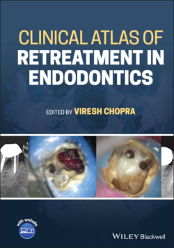
Clinical Atlas of Retreatment in Endodontics

Реклама. ООО «ЛитРес», ИНН: 7719571260.
Оглавление
Группа авторов. Clinical Atlas of Retreatment in Endodontics
Table of Contents
List of Illustrations
Guide
Pages
Clinical Atlas of Retreatment in Endodontics
Foreword
Foreword
Preface
Acknowledgments
List of Contributors
List of Abbreviations
About the Companion Website
Introduction to endodontic retreatment
I.1 Definition
I.2 Rationale for retreatment
I.3 Aim of endodontic retreatment
References
1 Clinical Case 1 – Perforation repair: A case of repair of pulpal floor perforation caused by excessive cutting of the floor of the pulp chamber
1.1 Patient information
1.2 Tooth
1.3 Treatment plan
1.4 Technical aspects
1.5 Follow‐up
1.6 Learning objectives
1.7 How can this endodontic mishap be avoided?
2 Clinical Case 2 – Instrument separation: A case of surgical removal of a fractured instrument
2.1 Patient information
2.2 Tooth
2.3 Treatment plan
2.4 Surgical procedure
2.5 Technical aspects
2.6 Follow‐up
2.7 Learning objectives. The reader should be able to:
3 Clinical Case 3 – A case of retreatment of Tooth 16: Bypass of ledges and broken instrument
3.1 Patient information
3.2 Tooth
3.3 Treatment plan
3.3.1 Shaping
3.3.2 Irrigation (solution and technique)
3.3.3 Obturation
3.4 Technical aspects
3.5 Follow‐up
3.6 Learning objectives
3.7 How can this endodontic mishap be avoided?
4 Clinical Case 4 – Instrument retrieval: A case of fractured instrument at the apical third of the mandibular molar
4.1 Patient information
4.2 Tooth
4.3 Treatment plan
4.4 Removing or bypassing the fractured instrument: decision making
4.5 Follow‐up
4.6 Learning objectives
4.7 How can this endodontic mishap be avoided?
5 Clinical Case 5 – Perforation repair with instrument retrieval: Management of multiple endodontic mishaps
5.1 Patient information
5.2 Tooth
5.3 Treatment plan
5.4 Technical aspects
5.5 Follow‐up
5.6 Learning objectives
5.7 How can this endodontic mishap be avoided?
6 Clinical Case 6 – Management of strip perforation and fractured instrument
6.1 Patient information
6.2 Tooth
6.3 Treatment plan
6.4 Technical aspects
6.5 Follow‐up
6.6 Learning objectives
6.7 How can this endodontic mishap be avoided?
7 Clinical Case 7 – Management of root canal treatment failure case with missed lateral canal anatomy and inadequate obturation
7.1 Patient information
7.2 Tooth
7.3 Treatment plan
7.4 Technical aspects
7.5 Follow‐up
7.6 Learning objectives
7.7 How can this endodontic mishap be avoided?
8 Clinical Case 8 – Management of a case with faulty cast post and asymptomatic lateral periodontitis
8.1 Patient information
8.2 Tooth
8.3 Treatment plan
8.4 Technical aspects
8.5 Follow‐up
8.6 Learning objectives
8.7 How can this endodontic mishap be avoided?
9 Clinical Case 9 – Management of a case with endo‐perio lesion following a previous root canal treatment
9.1 Patient information
9.2 Tooth
9.3 Treatment plan
9.4 Technical aspects
9.5 Follow‐up
9.6 Learning objectives
9.7 How can this endodontic mishap be avoided?
10 Clinical Case 10 – Management of a failed root canal treatment with silver cone obturation and fractured instrument
10.1 Patient information
10.2 Tooth
10.3 Treatment plan
10.4 Technical aspects
10.5 Follow‐up
10.6 Learning objectives
10.7 How can this endodontic mishap be avoided?
11 Clinical Case 11 – Management of a failed root canal treated maxillary molar with selective root treatment
11.1 Introduction to the concept of selective root treatment
11.2 Decision making
11.3 Laying down the treatment plan
11.4 Steps of selective retreatment
11.5 Introduction to the clinical case
11.5.1 Patient information
11.5.2 Tooth
11.5.3 Treatment plan
11.5.4 Technical aspects
11.5.5 Follow‐up
11.6 Conclusion
11.7 Learning objectives
11.8 How can this endodontic mishap be avoided?
References
12 Clinical Case 12 – Guided endodontics and its application for non‐surgical retreatments: Retreatment of a maxillary anterior tooth using static guidance
12.1 Introduction to guided endodontics
12.2 Pulp canal calcification
12.3 Improving the accuracy of access preparation
12.4 Static guidance
12.4.1 Advantages
12.4.2 Disadvantages
12.4.3 Challenges with static guidance
12.5 Approach to planning
12.5.1 Steps for planning and printing
12.6 Burs used for static guided endodontics
12.7 Introduction to the clinical case
12.7.1 Patient information
12.7.2 Tooth
12.7.3 Treatment plan
12.7.4 Technical aspects
12.7.5 Follow‐up
12.8 Learning objectives
12.9 How can this endodontic mishap be avoided?
12.10 FAQs for guided endodontics
12.11 Summary
References
13 Clinical Case 13 – Management of pulpal floor perforation with periapical lesion in the mesial root
13.1 Patient information
13.2 Tooth
13.3 Treatment plan
13.4 Technical aspects
13.5 Follow‐up
13.6 Learning objectives
13.7 How can this endodontic mishap be avoided?
14 Clinical Case 14 – Management of root canal treatment failure with missed canal anatomy and inadequate obturation
14.1 Patient information
14.2 Tooth
14.3 Treatment plan
14.4 Technical aspects
14.5 Follow‐up
14.6 Learning objectives
14.7 How can this endodontic mishap be avoided?
15 Clinical Case 15 – Management of root canal treatment failure with inadequate obturation, hidden fractured instrument and ledge formation in a severely curved mandibular molar
15.1 Patient information
15.2 Tooth
15.3 Treatment plan
15.4 Technical aspects
15.5 Follow‐up
15.6 Learning objectives
15.7 How can this endodontic mishap be avoided?
16 Clinical Case 16 – Management of root canal treatment with an instrument fracture in a mandibular molar
16.1 Patient information
16.2 Tooth
16.3 Treatment plan
16.4 Technical aspects
16.5 Follow‐up
16.6 Learning objectives
16.7 How can this endodontic mishap be avoided?
17 Clinical Case 17 – Management of a mandibular molar with fractured instrument extending in the periapical area
17.1 Patient information
17.2 Tooth
17.3 Treatment plan
17.4 Technical aspects
17.5 Learning objectives
17.6 How can this endodontic mishap be avoided?
18 Clinical Case 18 – Management of root canal treatment failure with inadequate obturation and apically calcified canals
18.1 Patient information
18.2 Tooth
18.3 Treatment plan
18.4 Technical aspects
18.5 Follow‐up
18.6 Learning objectives
18.7 How can this endodontic mishap be avoided?
19 Clinical Case 19 – Management of root canal treatment failure with inadequate obturation and missed canals
19.1 Patient information
19.2 Tooth
19.3 Treatment plan
19.4 Technical aspects
19.5 Follow‐up
19.6 Learning objectives
19.7 How can this endodontic mishap be avoided?
20 Clinical Case 20 – Management of root canal treatment failure with inadequate obturation, unusual distal root anatomy and suspected ledge formation in a mandibular molar
20.1 Patient information
20.2 Tooth
20.3 Treatment plan
20.4 Technical aspects
20.5 Follow‐up
20.6 Learning objectives
20.7 How can this endodontic mishap be avoided?
21 Clinical Case 21 – Management of root canal treatment failure with inadequate obturation and faulty post placement
21.1 Patient information
21.2 Tooth
21.3 Treatment plan
21.4 Technical aspects
21.5 Follow‐up
21.6 Learning objectives
21.7 How can this endodontic mishap be avoided?
22 Clinical Case 22 – Management of root canal treatment failure with inadequate obturation, multiple perforations, fractured instrument and ledge formation in maxillary right first molar
22.1 Patient information
22.2 Tooth
22.3 Treatment plan
The treatment was planned in different stages
22.4 Technical aspects
22.5 Follow‐up
22.6 Learning objectives. The reader should be able to understand:
22.7 How can this endodontic mishap be avoided?
23 Clinical Case 23 – Management of root canal treatment failure with inadequate obturation, fractured instrument and periapical lesion in mandibular left first molar
23.1 Patient information
23.2 Tooth
23.3 Treatment plan
23.4 Technical aspects
23.5 Follow‐up
23.6 Learning objectives. The reader should be able to understand:
23.7 How can this endodontic mishap be avoided?
24 Clinical Case 24 – Retreatment of Tooth 21
24.1 Patient information
24.2 Tooth
24.3 Treatment plan
24.4 Technical aspects
24.5 Follow‐up
24.6 Learning objectives
24.7 How can this endodontic mishap be avoided?
25 Nonsurgical versus surgical retreatment: Decision making
25.1 The evidence
25.2 The operator
25.3 The patient
25.4 Medical history
25.5 Medications
25.6 The tooth: factors to consider. Persistent periapical pathology
Presurgical CBCT evaluation
25.7 Quality of restoration and post
25.8 Root canal obturation quality and iatrogenic errors
25.9 Three clinical cases
Case 1 – preoperative microsurgical planning using CBCT imaging
Case 2 – decision for non‐surgical retreatment due to missed anatomy
Case 3 – decision for surgical treatment: PAP on mesial root only and no missed anatomy
References
Index
a
c
e
g
i
l
m
n
p
r
s
WILEY END USER LICENSE AGREEMENT
Отрывок из книги
Edited by
.....
I thank all the leading dental companies who trusted in this project and supported me for it. Thank you, Carl Zeiss, Zirc, FKG Dentaire, Coltène/Whaledent and bioMTA for your support.
I would like to thank the wonderful team at WILEY BLACKWELL for their genuine passion and professionalism for making this dream a reality. Thank you, Susan Engelken, Miss. Loan Nguyen, Tanya McMullin, Copyeditor Holly Regan‐Jones and Mustaq Ahamed for your support.
.....