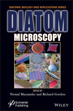
Diatom Microscopy

Реклама. ООО «ЛитРес», ИНН: 7719571260.
Оглавление
Группа авторов. Diatom Microscopy
Table of Contents
List of Illustrations
List of Tables
Guide
Pages
Diatom Microscopy
Preface
References
1. Investigation of Diatoms with Optical Microscopy
1.1 Introduction
1.2 Light Microscopy. 1.2.1 Phase Contrast Microscopy
1.2.2 Differential Interference Contrast (DIC) Microscopy
1.2.3 Darkfield Microscopy
1.3 Fluorescence Microscopy
1.4 Confocal Laser Scanning Microscopy
1.5 Multiphoton Microscopy
1.6 Super-Resolution Optical Microscopy
1.7 Conclusion
Acknowledgement
References
2. Nanobioscience Studies of Living Diatoms Using Unique Optical Microscopy Systems
Abbreviations
2.1 Trajectory Analysis of Gliding Among Individual Diatom Cells Using Microchamber Systems
2.2 Direct Observation of Floating Phenomena of Individual Diatoms Using a “Tumbled” Microscope System
2.3 Three-Dimensional Physical Imaging of Living Diatom Cells Using a Holographic Microscope System
Acknowledgements
References
3. Recent Insights Into the Ultrastructure of Diatoms Using Scanning and Transmission Electron-Microscopy
3.1 Introduction
3.2 Scanning Electron Microscopy (SEM) of Diatoms
3.3 Transmission Electron Microscopy (TEM) of Diatoms
3.3.1 Limitations
3.4 Conclusion
References
4. Atomic Force Microscopy Study of Diatoms
4.1 Introduction
4.2 Types of AFM Modes
4.3 Sample Preparation and Methods
4.4 Study of Diatom Ultrastructure Under AFM
4.5 Conclusion
Glossary
Acknowledgement
References
5. Refractive Index Tomography for Diatom Analysis
5.1 Introduction
5.2 Fundamentals of PC-ODT
5.3 Experimental Setup for PC-ODT
5.4 Diatom RI Reconstructions with Bright-Field Illumination
5.5 Illumination Impact on PC-ODT Performance
5.6 Concluding Remarks
Acknowledgement
References
6. Luminescent Diatom Frustules: A Review on the Key Research Applications
6.1 Introduction
6.2 Key Research Applications of Luminescence Properties of Diatom Frustules
6.2.1 Novel Nanophotonic and Optoelectronic Applications of Luminescent Diatom Frustules
6.2.2 Applications of Diatom Luminescence in Sensing
6.2.3 Biomedical Applications of Diatom Luminescence
6.2.4 Other Studies on Diatom Luminescence
6.3 Future Perspectives
6.4 Conclusion
Acknowledgement
References
7. Micro to Nano Ornateness of Diatoms from Geographically Distant Origins of the Globe
7.1 Introduction
7.2 Materials and Methods. 7.2.1 Diatom Samples and Microscopy. 7.2.1.1 By Michael J. Stringer
7.2.1.2 Diatom Oamaru Slides by Diane Winter
7.2.1.3 By Daniel Mathys
7.2.1.4 Diatom Sampling, Slide Preparation and Imaging from Himalayas, Plains and Arabian Sea, India
7.3 Diatoms from Different Geographical Origins of the World. 7.3.1 Oamaru Diatoms
7.3.2 Diatom Images Gifted by Michael J. Stringer
7.3.3 Diatoms from Natural History Museum Basel, Switzerland a Piece of Art by Daniel Mathys
7.3.4 Diatoms from India
7.4 Conclusion
7.5 Acknowledgements
References
8. Types of X-Ray Techniques for Diatom Research
8.1 Introduction
8.2 Applications. 8.2.1 Synchrotron Radiation-Based X-Ray Techniques
8.2.2 X-Ray Computed Tomography
8.2.3 X-Ray Fluorescence-Based Techniques
8.2.4 X-Ray Microanalysis
8.2.5 X-Ray Absorption-Based Techniques
8.2.6 X-Ray Diffraction
8.2.7 Other X-Ray-Based Techniques
8.3 Conclusions
Glossary
References
9. Diatom Assisted SERS
9.1 Introduction
9.2 Diatom. 9.2.1 Basic Overview
9.2.2 Physiological Characteristics
9.2.3 Optical and Relevant Properties
9.3 Raman Scattering. 9.3.1 Basics
9.3.2 Surface Enhanced Raman Scattering
9.3.3 Optoelectronic Investigations
9.4 SERS Through Diatom: Fundamentals and Application Overview
9.5 Conclusion and Future Outlook
References
10. Diatoms as Sensors and Their Applications
10.1 Introduction
10.2 Diatoms as Biosensors
10.2.1 Electrochemical Sensors
10.2.2 Plasmonic Sensors
10.2.3 Immunoassay Sensors
10.2.4 Optical and Optofluidic Sensors
10.2.5 Biochemical Sensors
10.2.6 FRET-Based Sensors
10.2.7 Microfluidics-Based Sensors
10.3 Conclusion
Acknowledgments
References
11. Diatom Frustules: A Transducer Platform for Optical Detection of Molecules
11.1 Introduction
11.2 Optical Properties of Diatom Frustules. 11.2.1 Diatom as a Photoluminescent Materials
11.2.2 Diatom as a Photonic Crystal
11.2.3 Diatoms as a SERS Substrate
11.3 Methods Involved in Thin Film Deposition of Diatom Frustules
11.4 Diatom as an Optical Transducer for Biosensors
11.5 Diatom as an Optical Transducer for Gas/Chemical Sensors
11.6 Conclusion
References
12. Effects of Light on Physico-Chemical Properties of Diatoms
12.1 Introduction
12.2 Effect of Light on Diatom Function and Morphology
12.2.1 Effect of Light Intensity on Diatom Morphology
12.2.2 Effect of Light Intensity on Diatom Growth
12.2.3 Effect of Light Intensity on Photosynthesis in Diatoms
12.2.4 Effect of Wavelength of Light on Diatom Pigment System
12.2.5 Effect of Light Intensity on the Physiology of Diatoms
12.3 Conclusion
Acknowledgment
References
Index
Also of Interest
WILEY END USER LICENSE AGREEMENT
Отрывок из книги
Scrivener Publishing 100 Cummings Center, Suite 541J Beverly, MA 01915-6106
.....
[1.12] Denk, W., Strickler, J.H. and Webb, W.W. (1990) Two-photon laser scanning fluorescence microscopy. Science 248(4951), 73–76.
[1.13] Dertinger, T., Colyer, R., Iyer, G., Weiss, S. and Enderlein, J. (2009) Fast, background-free, 3D super-resolution optical fluctuation imaging (SOFI). Proc Natl Acad Sci USA 106(52), 22287–22292.
.....