Читать книгу King's Applied Anatomy of the Abdomen and Pelvis of Domestic Mammals - Geoff Skerritt - Страница 26
1.4 The Sheath of the Rectus Abdominis Muscle (Figures 1.10a–c)
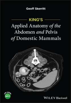
ОглавлениеFigure 1.10 Transverse sections through the ventral body wall to show the species variation in the sheath of the rectus abdominis. (a) The horse, (b) the ox and (c) the dog (caudal third of abdomen only).1 = parietal peritoneum; 2 = transverse abdominis muscle; 3 = interior oblique abdominis muscle; 4 = exterior oblique abdominis muscle; 5 = yellow abdominis tunic; 6 = skin; 7 = rectus abdominis muscle; 8 = linea alba; 9 = ventral sheath of rectus abdominis muscle
The aponeuroses of the external, internal and transverse abdominal oblique muscles together form a sheath that encloses the rectus abdominis muscle either side of the midline of the abdominal wall. There are species differences and, in the dog, variations in the craniocaudal location.
Species variations: In the caudal third of the abdomen of the dog the tendon of the transverse abdominal muscle lies ventral to the rectus abdominis muscle. In the middle third of the abdomen the transverse abdominal tendon lies dorsal, and that of the internal abdominal oblique muscle passes ventral to the rectus abdominis (as in the horse). In the cranial third of the abdomen the tendon of the transverse abdominal muscle lies dorsal to the rectus muscle. In addition, the internal oblique tendon divides into a ventral and dorsal portion (as in the ox).
In the horse the aponeurosis of the internal oblique muscle lies ventral to the rectus abdominis. In addition, in this species, the yellow abdominal tunic is present.
In the ox the aponeurosis of the internal oblique divides to pass on both sides of the rectus abdominis, and a yellow abdominal tunic is again present. In this species the linea alba is particularly wide.