Читать книгу Orthodontic Treatment of Impacted Teeth - Adrian Becker - Страница 80
3D module
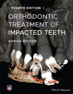
ОглавлениеA majority of dental CBCT software will have some kind of 3D volume‐rendering module, which is a very valuable tool for the accurate positional diagnosis and treatment planning of impacted teeth. The 3D volume‐rendering module depicts the individual teeth in their exact spatial arrangements and proximity to one another, from root apices to crown tips and viewable from every angle. The capabilities of this module vary between software programs and normally include several viewing modes. A logical examination sequence would start with the 3D rendering module, during which the ROI is identified, before moving on to slicing these areas.
It is possible to move and rotate the volume, to ‘sculpt’ away areas that interfere or obstruct, to clip in a given axis and to peel away bone. In relation to impacted teeth, the most popular viewing modes in the orthodontic context are the transparent mode and the opaque bony appearance. The 3D module is good for a general, overall survey and will help clarify the crown and root relationships of impacted and supernumerary teeth with adjacent structures. Unfortunately, 3D portrayal cannot be trusted to discern tooth contact or minor resorption, not even using ideal viewing angles. Bone peeling, relevant to the opaque bony viewing mode, is not a tooth segmentation procedure, because this will peel off visual information with a density below the set threshold. When cortical bone areas have a similar density to that of dentine, the software is unable to distinguish between the two and will peel both. When the 3D threshold control is altered, the visible tooth volume changes. Thus, when thicker cortical bone needs to be peeled off, more dentine will be peeled off with it and a smaller tooth volume will result.
