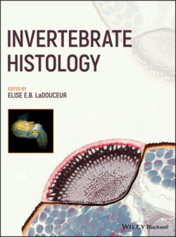
Читать книгу Invertebrate Histology - Группа авторов - Страница 18
1.3.3 Digestive System
ОглавлениеEchinoderms are a diverse group of animals with different nutritional strategies reflected in their digestive tracts. All consist of a simple tubular structure extending from the mouth to the anus with varying modifications that aid in digestion. In asteroids, the alimentary canal consists of a mouth, esophagus, stomach (cardiac, pyloric), intestine, and rectum. The mouth is at the center of the peristomial membrane and is separated by a muscular sphincter from the short esophagus and a more complex stomach. The cardiac portion of the stomach is large and has 10 distinct pouch‐like structures (radial pouches). Five of the pouches extend into the lumen of the arm from the disc and are attached to the ambulacral ossicles by muscle and dense connective tissue. A pair of gastric ligaments anchors the esophagus and permits retraction of the cardiac stomach in species that evert it during feeding. Above the radial pouches are five interradial pouches that eventually transition into the pyloric portion of the stomach. The pyloric stomach is smaller, flattened, and “star shaped” with five ducts that each extend into the central coelomic cavity of each ray and connect with the heavily branched pyloric cecae. The upper portion of the stomach tapers to form a short intestine that can have its own series of short blind sacs (intestinal cecae). The intestine connects to the short rectum and anus (Leake 1975; Ruppert et al. 2004).
The gastrodermis of the asteroid cardiac stomach is a pseudostratified columnar epithelium (Figure 1.17a). These cells lie on a basal lamina and basiepithelial nerve plexus with a connective tissue wall and outer coelomic epithelial liming. Circular and longitudinal muscle layers are interwoven into the coelomic lining. The gastrodermis is composed of supporting cells, secretory cells, and two types of coelomocytes. Supporting cells have a single cilium and numerous long microvilli. Secretory cells have no cilia and are either mucous or glandular in type. Two types of coelomocytes are normally seen in the gastrodermis and are found at all levels of the gut wall. The gastrodermis of the pyloric stomach is similar to the cardiac. Both the gastrodermal lining and the entire wall are thinner in the pyloric stomach due to reduced presence/thickness of the basiepithelial nerve layers, connective and muscle tissue layers (Figure 1.17b).
Figure 1.17 Histology of the cardiac (a) stomach in a mottled star (100×, HE) and pyloric stomach (b) of a mottled star (200×, HE).
Figure 1.18 Histology of the pyloric cecae of a mottled star. 25×, HE.
The pyloric and intestinal cecae are only present in asteroids. They are foliate structures created by extensive diverticula, which extend laterally from a medial duct. The diverticula are further divided into secondary chambers that are arranged parallel to the median duct. The lining of the pyloric cecae consists of very tall ciliated supporting cells and glandular secretory cells (mucous and zymogen cells) which are most abundant in the distal chambers of the pyloric cecae. Storage cells, cells containing large lipid vacuoles and polysaccharide and glycogen laden vacuoles, are more abundant distally (Hyman 1955; Leake 1975) (Figure 1.18).
The gastrodermis of the intestine and intestinal cecae is a ciliated pseudostratified columnar epithelium that in some areas may be compressed into a simple columnar epithelium and appear similar to the lining of the stomach. The epithelium is composed of supporting cells and two types of mucous secretory cells. The muscle, connective tissue, and nervous components are poorly developed in the intestinal cecal wall. The gastrodermis of the rectum and anus are identical and consist of a pseudostratified columnar epithelium composed predominantly of monociliated supporting cells attached to a basal lamina. The basiepithelial nerve plexus is reduced to absent. The connective tissue layer is thicker than in the intestine and pyloric cecae (5–10 μm thick) and is composed of thin elastic fibers (Hyman 1955). In Ophiurioidea, the digestive system is composed of a mouth, esophagus, stomach, rectum, and anus but lacks an intestinal tract and all components have histologic features similar to those described in asteroids.
In Echinoidea there is a mouth and a unique masticatory apparatus, Aristotle's lantern, followed by the esophagus, intestine, rectum, and anus. Aristotle's lantern is a pentamerous cone made of 40 ossicles including five teeth, adjoined by muscles and confined by coelomic membranes. At the ventrum of the lantern, the mouth is surrounded by a peristomial membrane, composed of mutable collagenous tissue covered in epidermis. Food passes through the mouth into a short pentagonal pharynx suspended in the center of the lantern. The pharynx transitions to esophagus at the top of the lantern. The esophagus ascends and then loops back as intestine. A blind pouch, variably referred to as stomach or cecum, may be present at the junction of esophagus and intestine. The intestine coils along the inside of the test, suspended by peritoneal membranes (i.e., mesenteries). The first nearly complete coil courses counterclockwise (when viewed from a dorsal or aboral aspect), and this segment is sometimes referred to as the stomach, or small or inferior intestine. Most echinoids have a slender extension of the intestine that accompanies this first coil at its inner border, termed the siphon, and it is believed to facilitate extraction of water from food. Then, the intestine turns back on itself and courses dorsally and clockwise to form a second coil, and this segment is sometimes referred to as the large or superior intestine. Finally, the terminal intestine forms the rectum that ascends to the interior of the periproct and forms the anus.
Histologically, the echinoid digestive tract has layers similar to other echinoderms. The epithelial lining is composed of tall columnar ciliated epithelial cells termed enterocytes, some of which bear microvilli, and others that may be distinguished as mucous cells (Figure 1.19). Similar to the epidermis, there is a subtle nervous layer at the base of enterocytes. Subjacent to this is a thin layer of connective tissue, followed by a thinner layer of muscle cells, typically arranged in a circular pattern relative to the lumen. The outer layer consists of a simple layer of flagellated cuboidal epithelial cells, as found on coelomic surfaces of other viscera. Glandular crypts may form where shortened enterocytes segmentally invaginate. Oral (small) and aboral (large) intestine may be histologically distinguished by differential presence of glands, villi, thickness, or prominence of microvilli (Work n.d.; Francis‐Floyd et al. 2020). The siphon is histologically similar to the small intestine, only of smaller diameter. Histologic sections through the lantern typically feature major ossicles (i.e., the pyramids, compass, and rotula), teeth, interpyramidal (or comminator) muscles, the pharynx, peristomial membrane, the circumoral nerve ring, and sometimes gill at the lateral margin of the lantern (Figure 1.20). The central cavity of the lantern coelom that surrounds the pharynx reflects between folds of interpyramidal muscle. Its myocytes are arranged into rows along a thin connective tissue septum and are covered by a layer of squamous and ciliated adluminal cells (Märkel et al. 1990). The protractor and retractor muscles exterior to the base of the lantern are instead arranged into fascicles within connective tissue matrix (Ziegler et al. 2012).
Figure 1.19 Histology of the large intestine of a white urchin. 200×, HE.
Figure 1.20 Low‐magnification histology of anatomy of Aristotle's lantern in a white urchin. Inset shows closer view of interpyramidal muscle. 20×, HE. I, interpyramidal muscle; M, mouth; P, pharynx; T, teeth.
In Holothuroidea, there is a mouth, pharynx (calcareous ring), esophagus, stomach, anterior and posterior intestine, and cloaca. The mouth is at the center of a buccal membrane and is surrounded by a muscular sphincter. This leads to a short pharynx enclosed in a ring of ossicles. The stomach may not be present in some species and is generally not as well defined as in Asteroidea. The pharynx and stomach have a tall columnar epithelial lining composed of supporting and glandular cells showing mucous cell differentiation. Both have an internal cuticular lining unlike other species. The intestinal tract in holothuroids is extensive and is the primary site of digestion. The anterior portion (small intestine) has an extensive associated vascular system. It is lined by tall ciliated epithelial cells with prominent glandular differentiation and has a thin muscular wall. The posterior portion (large intestine) has a thinner epithelium with more prominent mucous cell differentiation. The digestive system of Crinoidea is confined to the disc and consists of a mouth, esophagus, intestine, rectum, and anus (anal cone) (Ruppert et al. 2004). Histology is similar to previously described echinoderm species.