Читать книгу An Illustrated Guide to Oral Histology - Группа авторов - Страница 14
1 Tooth Development
ОглавлениеSaqib Ali1, Imran Farooq1, and Syed Ali Khurram2
1 Department of Biomedical Dental Sciences, College of Dentistry, Imam Abdulrahman Bin Faisal University, Dammam, Saudi Arabia
2 Unit of Oral and Maxillofacial Pathology, School of Clinical Dentistry, University of Sheffield, Sheffield, United Kingdom
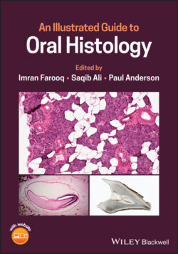
Figure 1.1 H and E stained section showing tooth development.
Tooth development starts on the 37th day of gestation with the formation of primary epithelial bands in the place of future upper and lower jaws. These horse‐shoe‐shaped bands correspond to the future dental arches. These epithelial bands then form two ingrowths called dental lamina (lingually positioned) and vestibular lamina (buccally positioned). These ingrowths extend into the mesenchyme which is surrounded by the neural crest cells. The vestibular lamina proliferates within the mesenchyme and leads to the formation of the vestibule (between the cheek and tooth‐bearing portion of the jaw). The dental lamina gives rise to epithelial outgrowths toward the mesenchyme due to continuous proliferative activity which correspond to the location of forthcoming deciduous teeth. The tooth development is divided into the following stages: bud, cap, and bell (early and late). These stages along with the changes happening in the tooth germ are discussed in detail in the following sections.