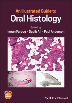
Читать книгу An Illustrated Guide to Oral Histology - Группа авторов - Страница 2
Table of Contents
Оглавление1 Cover
2 Title Page
3 Copyright Page
4 Preface
5 Sample Preparation Hematoxylin and Eosin (H and E) Stained Sections Micro‐computed Tomography (Micro‐CT) Ground Sections Scanning Electron Microscopy
6 About the Editors
7 List of Contributors
8 About the Companion Website
9 1 Tooth Development 1.1 Bud Stage 1.2 Cap Stage 1.3 Early Bell Stage 1.4 Late Bell Stage 1.5 Root Formation 1.6 Amelogenesis Imperfecta (AI) 1.7 Dentinogenesis Imperfecta (DI) 1.8 Dentin Dysplasia (DD) References
10 2 Dental Enamel 2.1 Surface Enamel and Ionic Substitution 2.2 Enamel Striae 2.3 Enamel Lamellae 2.4 Enamel Spindles 2.5 Enamel Tufts 2.6 Enamel Dentin Junction (EDJ) 2.7 Neonatal Line 2.8 Gnarled Enamel 2.9 Enamel Caries References
11 3 Dentin 3.1 Dentinal Tubules 3.2 Organic Matrix of Dentin 3.3 Primary and Secondary Curvatures of Tubules 3.4 Interglobular Dentin 3.5 Peritubular/Intratubular and Intertubular Dentin 3.6 Dead Tracts 3.7 Tertiary Dentin 3.8 Sclerotic Dentin 3.9 Tome's Granular Layer (TGL) 3.10 Dentin Caries References
12 4 Cementum 4.1 Acellular Cementum 4.2 Cellular Cementum 4.3 Cementocytes and Lacunae 4.4 Cementoenamel Junction (CEJ) 4.5 Hypercementosis 4.6 Cementoblastoma 4.7 Root Resorption References
13 5 Dental Pulp 5.1 Odontogenic Zone 5.2 Cell‐Free Zone of Weil 5.3 Cell‐Rich Zone 5.4 Pulp Core 5.5 Pulpal Fibrosis 5.6 Pulp Stones 5.7 Periapical Granuloma References
14 6 Periodontal Ligament 6.1 Gingival Fibers 6.2 Transseptal Fibers 6.3 Alveolar Crest Fibers 6.4 Horizontal Fibers 6.5 Oblique Fibers 6.6 Apical Fibers 6.7 Interradicular Fibers 6.8 Gingivitis 6.9 Periodontitis References
15 7 Alveolar Bone 7.1 Compact Bone 7.2 Circumferential Lamellae 7.3 Concentric Lamellae 7.4 Interstitial Lamellae 7.5 Osteocytes and Lacunae 7.6 Haversian Canals 7.7 Volkmann's Canals 7.8 Osteons 7.9 Spongy Bone 7.10 Marrow Spaces 7.11 Osteoporosis 7.12 Osteomyelitis 7.13 Osteoma 7.14 Osteitis Deformans (Paget's Disease) 7.15 Osteosarcoma References
16 8 Oral Mucosa 8.1 Fungiform Papillae 8.2 Filiform Papillae 8.3 Circumvallate Papilla 8.4 Taste Buds 8.5 Keratinized Oral Epithelium 8.6 Parakeratinized Oral Epithelium 8.7 Non‐Keratinized Oral Epithelium 8.8 Non‐Specific Ulcer 8.9 Oral Lichen Planus 8.10 Pemphigoid 8.11 Lipoma 8.12 Oral Epithelial Dysplasia 8.13 Oral Melanoma References
17 9 Salivary Glands 9.1 Serous Salivary Gland 9.2 Mucous Salivary Gland 9.3 Seromucous (Mixed) Salivary Gland 9.4 Intercalated Ducts 9.5 Striated Ducts 9.6 Excretory Ducts 9.7 Sialadenitis 9.8 Necrotizing Sialometaplasia 9.9 Pleomorphic Adenoma 9.10 Warthin Tumor References
18 Index
19 End User License Agreement
