Читать книгу Applied Oral Physiology - Robin Wilding - Страница 46
На сайте Литреса книга снята с продажи.
3.5.1 Periodontal Ligament Fibers
ОглавлениеThe periodontal ligament fibers are mainly collagen with some reticulin, elastin, and oxytalan fibers. Many of the collagen fibers are gathered together in bundles (the so-called principal fibers). These fiber bundles have been divided into groups on the basis of their direction and site. We can recognize apical, oblique, horizontal, alveolar crest, interradicular, and transalveolar fibers (▶ Fig. 3.7). It should be recalled that the fibers of the gingiva also contribute to the collagen fibers of the periodontal ligament. They ensure the firm but resilient attachment of the gingiva and teeth to the alveolar bone.
Oxytalan fibers are unlike collagen in that they are not banded, but they do consist of fibrils running parallel to the long axis of the fiber. They are more numerous nearer the tooth than the alveolar bone. They seem to be more numerous in teeth which are under heavy loads such as those supporting fixed partial prosthesis. The means whereby oxytalan fibers contribute to tooth support are controversial, but their association with blood vessels has led to the suggestion that they may maintain patency of vessels even during the moments of compression of the ligament.
The elastin fibers are confined to the walls of the blood vessels and the reticulin fibers to basement membranes.
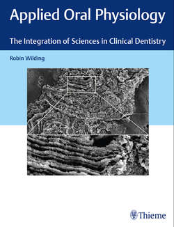
Fig. 3.7 SEM images of the two separate areas of periodontal ligament remaining attached to an extracted tooth (magnification × 2,000). (a) The collagen fiber bundles in this area of the PDL are orientated between the cementum surface (C) and the outer surface of the ligament which has separated from the tooth socket, estimated by the broken line. (b) The fiber bundles in this area of the periodontal ligament (PDL) are longer and appear to run in an oblique direction.