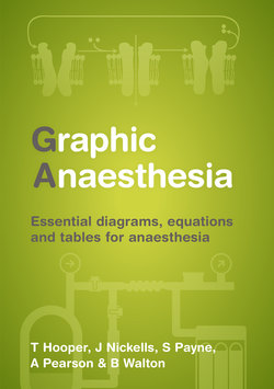
Читать книгу Graphic Anaesthesia - Tim Hooper - Страница 6
На сайте Литреса книга снята с продажи.
ОглавлениеContents
Preface
About the authors
Abbreviations
SECTION 1 PHYSIOLOGY
| 1.1 | Cardiac | ||
| 1.1.1 | Cardiac action potential – contractile cells | ||
| 1.1.2 | Cardiac action potential – pacemaker cells | ||
| 1.1.3 | Cardiac action potential – variation in pacemaker potential | ||
| 1.1.4 | Cardiac cycle | ||
| 1.1.5 | Cardiac output equation | ||
| 1.1.6 | Central venous pressure waveform | ||
| 1.1.7 | Central venous pressure waveform – abnormalities | ||
| 1.1.8 | Einthoven triangle | ||
| 1.1.9 | Ejection fraction equation | ||
| 1.1.10 | Electrocardiogram | ||
| 1.1.11 | Electrocardiogram – cardiac axis and QTc | ||
| 1.1.12 | Fick method for cardiac output studies | ||
| 1.1.13 | Frank–Starling curve | ||
| 1.1.14 | Oxygen flux | ||
| 1.1.15 | Pacemaker nomenclature – antibradycardia | ||
| 1.1.16 | Pacemaker nomenclature – antitachycardia (implantable cardioverter defibrillators) | ||
| 1.1.17 | Preload, contractility and afterload | ||
| 1.1.18 | Pulmonary artery catheter trace | ||
| 1.1.19 | Systemic and pulmonary pressures | ||
| 1.1.20 | Valsalva manoeuvre | ||
| 1.1.21 | Valsalva manoeuvre – clinical applications and physiological abnormalities | ||
| 1.1.22 | Vaughan–Williams classification | ||
| 1.1.23 | Ventricular pressure–volume loop – left ventricle | ||
| 1.1.24 | Ventricular pressure–volume loop – right ventricle | ||
| 1.2 | Circulation | ||
| 1.2.1 | Blood flow and oxygen consumption of organs | ||
| 1.2.2 | Blood vessel structure | ||
| 1.2.3 | Hagen–Poiseuille equation | ||
| 1.2.4 | Laminar and turbulent flow | ||
| 1.2.5 | Laplace’s law | ||
| 1.2.6 | Ohm’s law | ||
| 1.2.7 | Starling forces in capillaries | ||
| 1.2.8 | Starling forces in capillaries – pathology | ||
| 1.2.9 | Systemic vascular resistance | ||
| 1.3 | Respiratory | ||
| 1.3.1 | Alveolar gas equation | ||
| 1.3.2 | Alveolar partial pressure of oxygen and blood flow | ||
| 1.3.3 | Bohr equation | ||
| 1.3.4 | Carbon dioxide dissociation curve and Haldane effect | ||
| 1.3.5 | Closing capacity | ||
| 1.3.6 | Dead space and Fowler’s method | ||
| 1.3.7 | Diffusion | ||
| 1.3.8 | Dynamic compression of airways | ||
| 1.3.9 | Fick principle and blood flow | ||
| 1.3.10 | Forced expiration curves | ||
| 1.3.11 | Functional residual capacity of the lungs | ||
| 1.3.12 | Lung and chest wall compliance | ||
| 1.3.13 | Lung pressure–volume loop | ||
| 1.3.14 | Lung volumes and capacities | ||
| 1.3.15 | Oxygen cascade | ||
| 1.3.16 | Oxygen dissociation curve and Bohr effect | ||
| 1.3.17 | Pulmonary vascular resistance | ||
| 1.3.18 | Pulmonary vascular resistance and lung volumes | ||
| 1.3.19 | Respiratory flow–volume loops | ||
| 1.3.20 | Shunt | ||
| 1.3.21 | Ventilation–perfusion ratio | ||
| 1.3.22 | Ventilatory response to carbon dioxide | ||
| 1.3.23 | Ventilatory response to oxygen | ||
| 1.3.24 | West lung zones | ||
| 1.3.25 | Work of breathing | ||
| 1.4 | Neurology | ||
| 1.4.1 | Action potential | ||
| 1.4.2 | Cerebral blood flow and blood pressure | ||
| 1.4.3 | Cerebral blood flow variation with ventilation | ||
| 1.4.4 | Cerebrospinal fluid | ||
| 1.4.5 | Gate control theory of pain | ||
| 1.4.6 | Glasgow Coma Scale | ||
| 1.4.7 | Intracranial pressure–volume relationship | ||
| 1.4.8 | Intracranial pressure waveform | ||
| 1.4.9 | Neuron | ||
| 1.4.10 | Neurotransmitters – action | ||
| 1.4.11 | Neurotransmitters – classification | ||
| 1.4.12 | Reflex arc | ||
| 1.4.13 | Synaptic transmission | ||
| 1.4.14 | Types of nerve | ||
| 1.4.15 | Visual pathway | ||
| 1.5 | Renal | ||
| 1.5.1 | Autoregulation of renal blood flow | ||
| 1.5.2 | Clearance | ||
| 1.5.3 | Glomerular filtration rate | ||
| 1.5.4 | Loop of Henle | ||
| 1.5.5 | Nephron | ||
| 1.5.6 | Renin–angiotensin–aldosterone system | ||
| 1.6 | Gut | ||
| 1.6.1 | Bile | ||
| 1.6.2 | Mediators of gut motility | ||
| 1.7 | Acid–base | ||
| 1.7.1 | Acid–base disturbances | ||
| 1.7.2 | Anion gap | ||
| 1.7.3 | Buffer solution | ||
| 1.7.4 | Dissociation constant and pKa | ||
| 1.7.5 | Henderson–Hasselbalch equation | ||
| 1.7.6 | Lactic acidosis | ||
| 1.7.7 | pH | ||
| 1.7.8 | Strong ion difference | ||
| 1.8 | Metabolic | ||
| 1.8.1 | Krebs cycle | ||
| 1.8.2 | Liver lobule | ||
| 1.8.3 | Nutrition and energy | ||
| 1.8.4 | Vitamins – sources and function | ||
| 1.8.5 | Vitamins – toxicity and deficiency | ||
| 1.9 | Endocrine | ||
| 1.9.1 | Adrenal gland | ||
| 1.9.2 | Adrenergic receptor actions | ||
| 1.9.3 | Catecholamine synthesis | ||
| 1.9.4 | Hypothalamic–pituitary–adrenal axis – anatomy | ||
| 1.9.5 | Hypothalamic–pituitary–adrenal axis – hormones | ||
| 1.9.6 | Vitamin D synthesis | ||
| 1.10 | Body fluids | ||
| 1.10.1 | Body fluid composition | ||
| 1.10.2 | Fluid compartments | ||
| 1.10.3 | Intravenous fluid composition | ||
| 1.11 | Haematology | ||
| 1.11.1 | Antibody | ||
| 1.11.2 | Blood groups | ||
| 1.11.3 | Coagulation – cascade (classic) model | ||
| 1.11.4 | Coagulation – cell-based model | ||
| 1.11.5 | Complement cascade | ||
| 1.11.6 | Haemoglobin | ||
| 1.11.7 | Prostanoid synthesis | ||
| 1.12 | Cellular | ||
| 1.12.1 | Cell | ||
| 1.12.2 | Cell membrane | ||
| 1.12.3 | G-proteins | ||
| 1.12.4 | Ion channels | ||
| 1.12.5 | Sodium/potassium–ATPase pump | ||
| 1.13 | Muscle | ||
| 1.13.1 | Actin–myosin cycle | ||
| 1.13.2 | Golgi tendon organ | ||
| 1.13.3 | Muscle spindle | ||
| 1.13.4 | Muscle types | ||
| 1.13.5 | Neuromuscular junction | ||
| 1.13.6 | Sarcomere | ||
| 1.13.7 | Skeletal muscle structure |
SECTION 2 ANATOMY
| 2.1 | Abdominal wall | ||
| 2.2 | Antecubital fossa | ||
| 2.3 | Autonomic nervous system | ||
| 2.4 | Base of skull | ||
| 2.5 | Brachial plexus | ||
| 2.6 | Bronchial tree | ||
| 2.7 | Cardiac vessels – cardiac veins | ||
| 2.8 | Cardiac vessels – coronary arteries | ||
| 2.9 | Circle of Willis | ||
| 2.10 | Cranial nerves | ||
| 2.11 | Cross-section of neck at C6 | ||
| 2.12 | Cross-section of spinal cord | ||
| 2.13 | Dermatomes | ||
| 2.14 | Diaphragm | ||
| 2.15 | Epidural space | ||
| 2.16 | Femoral triangle | ||
| 2.17 | Fetal circulation | ||
| 2.18 | Intercostal space | ||
| 2.19 | Internal jugular vein | ||
| 2.20 | Laryngeal innervation | ||
| 2.21 | Larynx | ||
| 2.22 | Limb muscle innervation (myotomes) | ||
| 2.23 | Lumbar plexus | ||
| 2.24 | Nose | ||
| 2.25 | Orbit | ||
| 2.26 | Rib | ||
| 2.27 | Sacral plexus | ||
| 2.28 | Sacrum | ||
| 2.29 | Spinal nerve | ||
| 2.30 | Thoracic inlet and first rib | ||
| 2.31 | Vertebra |
SECTION 3 PHARMACODYNAMICS AND KINETICS
| 3.1 | Clearance | ||
| 3.2 | Compartment model – one and two compartments | ||
| 3.3 | Compartment model – three compartments | ||
| 3.4 | Dose–response curves | ||
| 3.5 | Elimination | ||
| 3.6 | Elimination kinetics | ||
| 3.7 | Half-lives and time constants | ||
| 3.8 | Meyer–Overton hypothesis | ||
| 3.9 | Volume of distribution | ||
| 3.10 | Wash-in curves for volatile agents |
SECTION 4 DRUGS
| 4.1 | Anaesthetic agents – etomidate | ||
| 4.2 | Anaesthetic agents – ketamine | ||
| 4.3 | Anaesthetic agents – propofol | ||
| 4.4 | Anaesthetic agents – thiopentone | ||
| 4.5 | Local anaesthetics – mode of action | ||
| 4.6 | Local anaesthetics – properties | ||
| 4.7 | Neuromuscular blockers – mode of action | ||
| 4.8 | Neuromuscular blocking agents – depolarizing | ||
| 4.9 | Neuromuscular blocking agents – non-depolarizing | ||
| 4.10 | Opioids – mode of action | ||
| 4.11 | Opioids – properties | ||
| 4.12 | Volatile anaesthetic agents – mode of action | ||
| 4.13 | Volatile anaesthetic agents – physiological effects | ||
| 4.14 | Volatile anaesthetic agents – properties |
SECTION 5 PHYSICS
| 5.1 | Avogadro’s law | ||
| 5.2 | Beer–Lambert law | ||
| 5.3 | Critical temperatures and pressure | ||
| 5.4 | Diathermy | ||
| 5.5 | Doppler effect | ||
| 5.6 | Electrical safety | ||
| 5.7 | Electricity | ||
| 5.8 | Exponential function | ||
| 5.9 | Fick’s law of diffusion | ||
| 5.10 | Gas laws – Boyle’s law | ||
| 5.11 | Gas laws – Charles’ law | ||
| 5.12 | Gas laws – Gay-Lussac’s (Third Perfect) law | ||
| 5.13 | Gas laws – ideal gas law and Dalton’s law | ||
| 5.14 | Graham’s law | ||
| 5.15 | Heat | ||
| 5.16 | Henry’s law | ||
| 5.17 | Humidity | ||
| 5.18 | Laser | ||
| 5.19 | Metric prefixes | ||
| 5.20 | Power | ||
| 5.21 | Pressure | ||
| 5.22 | Raman effect | ||
| 5.23 | Reflection and refraction | ||
| 5.24 | SI units | ||
| 5.25 | Triple point of water and phase diagram | ||
| 5.26 | Types of flow | ||
| 5.27 | Wave characteristics | ||
| 5.28 | Wheatstone bridge | ||
| 5.29 | Work |
SECTION 6 CLINICAL MEASUREMENT
| 6.1 | Bourdon gauge | ||
| 6.2 | Clark electrode | ||
| 6.3 | Damping | ||
| 6.4 | Fuel cell | ||
| 6.5 | Monitoring of neuromuscular blockade | ||
| 6.6 | Oximetry – paramagnetic analyser | ||
| 6.7 | pH measuring system | ||
| 6.8 | Pulse oximeter | ||
| 6.9 | Severinghaus carbon dioxide electrode | ||
| 6.10 | Temperature measurement | ||
| 6.11 | Thermocouple and Seebeck effect |
SECTION 7 EQUIPMENT
| 7.1 | Bag valve mask resuscitator | ||
| 7.2 | Breathing circuits – circle system | ||
| 7.3 | Breathing circuits – Mapleson’s classification | ||
| 7.4 | Cleaning and decontamination | ||
| 7.5 | Continuous renal replacement therapy – extracorporeal circuit | ||
| 7.6 | Continuous renal replacement therapy – modes | ||
| 7.7 | Gas cylinders | ||
| 7.8 | Humidifier | ||
| 7.9 | Oxygen delivery systems – Bernoulli principle and Venturi effect | ||
| 7.10 | Piped gases | ||
| 7.11 | Scavenging | ||
| 7.12 | Vacuum-insulated evaporator | ||
| 7.13 | Vaporizer | ||
| 7.14 | Ventilation – pressure-controlled | ||
| 7.15 | Ventilation – volume-controlled |
SECTION 8 STATISTICS
| 8.1 | Mean, median and mode | ||
| 8.2 | Normal distribution | ||
| 8.3 | Number needed to treat | ||
| 8.4 | Odds ratio | ||
| 8.5 | Predictive values | ||
| 8.6 | Sensitivity and specificity | ||
| 8.7 | Significance tests | ||
| 8.8 | Statistical variability | ||
| 8.9 | Type I and type II errors |
