Читать книгу Haematology - Barbara J. Bain, Irene Roberts - Страница 20
Оглавление11 Pure erythroid leukaemia
A 40‐year‐old, previously fit man presented with progressive fatigue. Physical examination showed pallor and mild icterus. His full blood count showed Hb 87 g/l, WBC 1.7 × 10⁹/l, neutrophils 0.8 × 10⁹/l and platelets 26 × 109/l. His blood film showed no specific features but, importantly, red cell fragments, spherocytes and blast cells were not seen. Serum bilirubin and LDH were both mildly elevated. A direct Coombs test was negative and no auto‐ or allo‐antibodies were identified. He had a significantly raised haemoglobin F at 10.9%. The bone marrow aspirate and trephine biopsy sections were reviewed by the haematologists at the source hospital and a working diagnosis of myelodysplastic syndrome with haemolysis was proposed. Flow cytometric studies on the marrow aspirate, gating on the largest cells based on the forward scatter/side scatter (FSC/SSC) profile, identified CD34+ cells at 2% of events, whilst CD117+ cells were increased at 12% of events. The karyotype was normal. The patient was discussed at the regional multidisciplinary team meeting where the diagnosis was questioned and a central haematopathology review was recommended.
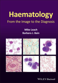
On review of the very cellular bone marrow aspirate, marked erythroid hyperplasia was evident (M:E ratio 1:10) with a notable left shift in erythroid precursors, prominent proerythroblasts, relatively mild erythroid dysplasia and karyorrhectic forms (top images ×100 objective). There was no excess of myeloblasts but dysplastic neutrophils were present (top centre). Importantly, the marrow was not megaloblastic. The trephine biopsy sections showed 100% cellularity with marked erythroid hyperplasia with left shift (bottom left, H&E and bottom centre, glycophorin C, immunoperoxidase ×50). A proportion of proerythroblasts also showed expression of CD117 (bottom right, immunoperoxidase ×50). There was no excess of CD34+ cells. A diagnosis of pure erythroid leukaemia was made. Subsequent myeloid next generation sequencing identified an NRAS mutation (c.35G>A) and two mutations in WT1 (c.758A>G and c.759C>A).
Pure erythroid leukaemia is a rare neoplastic bone marrow disorder characterised by a proliferation of immature erythroid cells (>80% of bone marrow cells with at least 30% proerythroblasts) and no significant myeloblast component. Patients typically present with pancytopenia and the condition can appear de novo, can evolve from a myelodysplastic syndrome or can be therapy‐related. It is important that the erythroid hyperplasia is not attributed to haemolysis (the evidence in this patient was not convincing) or to a myelodysplastic syndrome. In the latter, erythroid hyperplasia and dysplasia is common but the marked left shift with predominance of proerythroblasts and myeloid hypoplasia is not seen. Furthermore, de novo pure erythroid leukaemia, as in this 40‐year‐old patient, has a more acute presentation rather than the more gradual onset of cytopenias typically associated with MDS. A reversion to primitive erythropoiesis with increased haemoglobin F levels can occur in erythroleukaemia. There is no specific cytogenetic abnormality associated with pure erythroid leukaemia; complex karyotypes with loss of chromosomes 5 and 7, 5q− and 7q− are common, whilst favourable cytogenetics is very rare. The prognosis is poor when the condition evolves from MDS or is therapy‐related (these cases being categorised differently), but may be more favourable and similar to other subtypes of AML if it arises as a primary condition (Santos et al. 2009).
Reference
1 Santos FPS, Faderl S, Garcia‐Manero G, Koller C, Beran M, O’Brien S et al. (2009) Adult acute erythroid leukaemia: an analysis of 91 patients treated at a single institution. Leukemia, 23, 2275–2280.
MCQ
1 Proerythroblasts in pure erythroid leukaemia often express:CD34CD61CD117E‐cadherin (CD234)Glycophorin A (CD235a)For answers and discussion, see page 206.
