Читать книгу Dental Neuroimaging - Chia-shu Lin - Страница 36
1.2.4.1 T 1‐Weighted Structural MRI
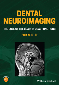
ОглавлениеThe most common sMRI data is acquired by T1‐weighted imaging. Imaging is acquired by weighing on the ‘T1’ value, which refers to the time constant for longitudinal relaxation, an index of the rate for protons to return to equilibrium. Critically, this value varies depending on the biochemical components of tissues: the fat‐containing tissue (e.g. neural fibres of white matter) has a shorter T1 compared to the tissue less rich in fat (e.g. grey matter). In a T1‐weighted image, the signals collected would preferably show a higher intensity (i.e. brighter) for brain tissue with a higher content of fat and lower intensity for tissue with a lower content of fat. Therefore, the spatial distribution of white matter and grey matter can be differentiated by image intensity. The greatest advantage of T1‐weighted imaging is that it can be analyzed using different methods to disclose information on brain morphology. For example, tissue‐specific segmentation is a method that separates grey matter, white matter and the space of CSF from the whole‐brain image. Voxel‐based morphometry (VBM) can be used to estimate the amount of grey matter and white matter within each voxel, which can be further used for a group‐based comparison. The method has been widely used for clinical investigation, such as assessing the reduction in hippocampal grey matter between patients with Alzheimer's disease and healthy controls. In addition, the surface‐based analysis is widely used to estimate the thickness of the cortical tissues and the volume of cortical and subcortical regions, which are critical structural features associated with clinical factors (see Section 2.3).
